Pelvic organ prolapse
Exam relevance: in 31 of 197 board exam reports · rank 25
- Synonyms
- uterine prolapse, genital prolapse
- Specialty
- Gynaecology · Pelvic floor & urogynaecology
- Images
- Colposcopy 1 · Clinical 4
- Exam relevance
- 31 of 197 reports · rank 25
- In the app
- 1 flashcards · GynFuchs
- Last updated
- 10/2026 · Dr. Pascal Bafteh
Contents
Images (5)
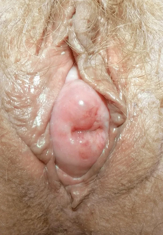 Colposcopy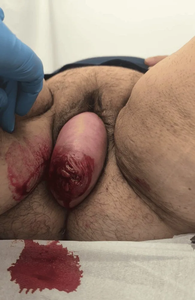
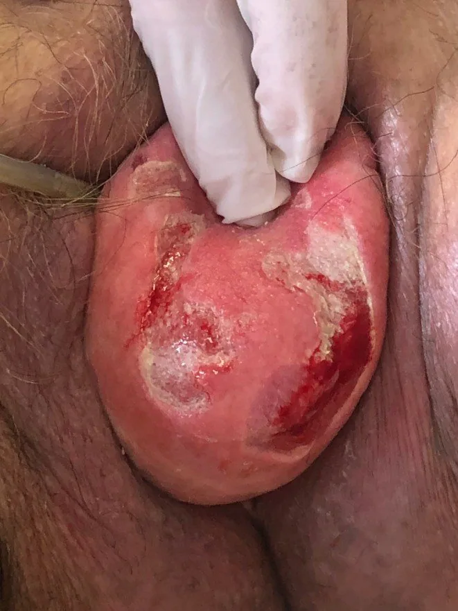
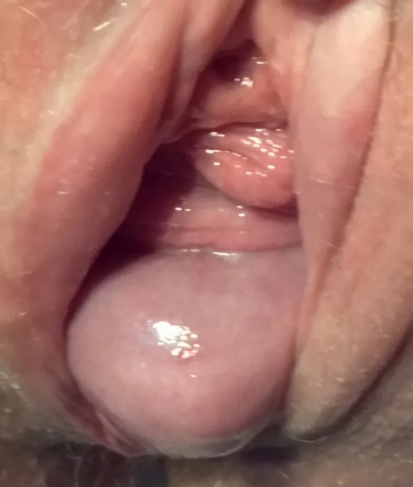
Classification
Three compartments
- Anterior compartment: cystocele and urethrocele, i.e. the anterior vaginal wall with the bladder and urethra.
- Posterior compartment: rectocele and enterocele with contents of the pouch of Douglas.
- Mnemonic for the examination: assess each compartment individually, as descensus often occurs in combination.
Grading
- Common clinical practice: grade I to III according to the position of the lowest point relative to the hymenal ring.
- Grade I: the lowest point remains above the hymenal ring.
- Grade II: the lowest point reaches approximately the introitus.
- Grade III: the descent protrudes beyond the introitus; a total prolapse is called procidentia.
- The differentiated system: POP-Q with six defined points, measured in centimetres relative to the hymen.
Clinical features
Symptoms and long-term consequences
- Key symptoms: foreign body and dragging sensation, low back pain and a pulling-down feeling.
- In severe cases: post-void residual volume, micturition and defecation disorders requiring manual reduction.
- Acute presentation can include: urinary retention, pain and bleeding from a decubitus ulcer of the prolapsed wall.
More facts from the study questions
- The clinical grading from I to III is based on the relationship of the lowest point to the hymenal ring.
- In Grade I, the lowest point of the prolapse is still located above the hymenal ring.
- The strength of the pelvic floor muscles is assessed as part of the clinical examination.
- Muscle strength is graded by palpation using the Oxford scale (Grades 0 to 5).
- The standard examination is performed in a supine position, with the patient asked to strain or cough.
- Genital prolapse is divided into three compartments: anterior, middle, and posterior.
- The anterior compartment comprises the anterior vaginal wall with the adjacent bladder (cystocele) and urethra (urethrocele).
- Grade II prolapse is present when the lowest point reaches approximately the introitus.
- If the prolapse extends beyond the introitus, it is a Grade III prolapse (or procidentia in case of total prolapse).
Diagnosis
Examination itself
- Examination is performed: in a supine position with straining and, if there is a discrepancy with the patient's complaints, additionally in a standing position.
- Without this step, the finding is underestimated: asking the patient to strain forcefully or cough.
- Bladder filling: the finding is re-evaluated with an empty bladder because it presents differently then.
- Pelvic floor strength: is graded on palpation from 0 to 5 using the Oxford scale.
Typical imaging findings
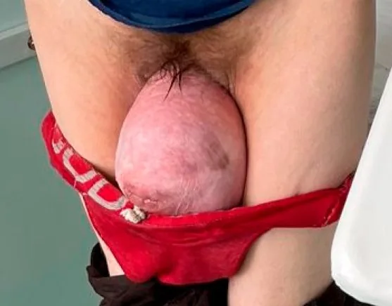
- Clinical finding: A prolapsed soft tissue body lies in front of the introitus. On its apex there are broad, sharply outlined defects with a fibrin-covered base and fresh bleeding points; the surrounding mucosa is dry and keratinised.
Keep learning in the app
Further reading (selection)
Cross-references
Note: Learning content from the GynFuchs app (flashcards, exam questions, image cases) for medical education – not a treatment recommendation and no substitute for diagnosis or treatment decisions in individual cases. Treatment and management are deliberately not covered on this page.