Uterine prolapse
Exam relevance: in 8 of 197 board exam reports · rank 82
- Specialty
- Gynaecology · Pelvic floor & urogynaecology
- Images
- Clinical 3 · Colposcopy 1
- Exam relevance
- 8 of 197 reports · rank 82
- Last updated
- 10/2026 · Dr. Pascal Bafteh
Contents
Images (4)
 Colposcopy
Definition
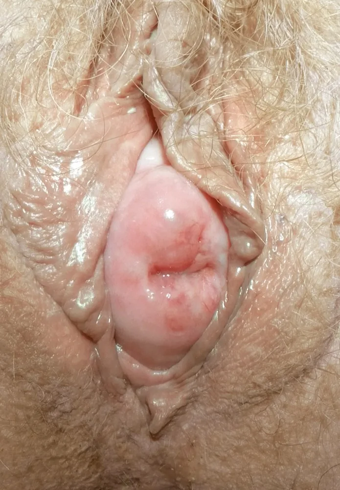
- Descent of the uterus with the cervix towards or past the introitus; in women without a uterus the vaginal vault (cuff) can descend in the same way (apical prolapse).
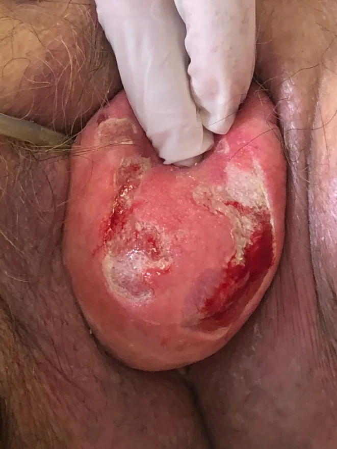
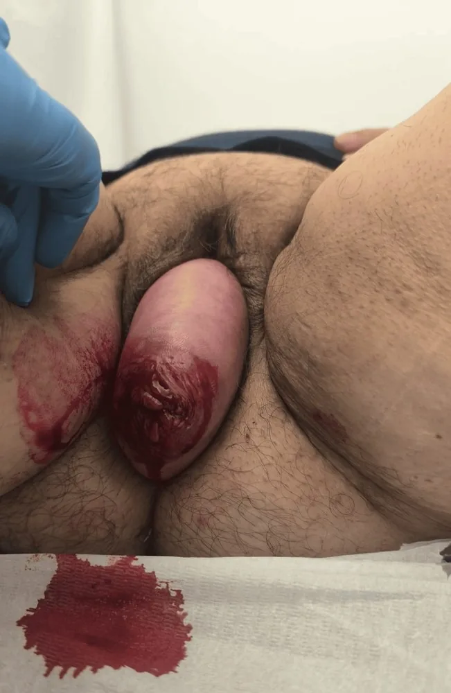
- In complete uterine prolapse (procidentia), the anterior and posterior vaginal walls collapse together with the apex, and the uterus may protrude out of the introitus.
Classification
- In the POP-Q system, point C describes the cervix or vaginal cuff and point D the posterior fornix; without prolapse the cervix lies about 4.5 to 7.5 cm above the hymenal remnants.
- POP-Q stages range from 0 (no prolapse) to IV (complete eversion); the older Baden-Walker system is considered imprecise and is no longer commonly used.
Occurrence & epidemiology
Epidemiology
- Pelvic organ prolapse is a common gynaecological condition, although reported prevalence varies; in a study of 8000 women, 8.3% reported symptomatic prolapse.
- A cystocele or rectocele is usually also present in uterine prolapse.
Aetiopathogenesis
Aetiology and pathogenesis
- It results from hernia-like laxity of the ligaments, fascia and muscles that support the pelvic organs.
- Common risk factors are vaginal births, particularly with a prolonged second stage, instrumental vaginal birth or a large newborn, as well as obesity, increasing age and chronically raised intra-abdominal pressure; sacral nerve and connective tissue disorders are less common.
Clinical features
- Symptoms tend to be minimal in mild prolapse; the most common presenting symptom is a vaginal bulge, which may be intermittent because of spontaneous reduction.
- In more severe prolapse, fullness, pressure, sexual dysfunction, a sensation of organs falling out, lower back pain, incomplete bladder emptying and constipation occur.
- Urinary incontinence is common; the descending organs may intermittently obstruct urine flow, causing urinary retention and overflow incontinence and masking stress incontinence.
- Mucosa prolapsed beyond the introitus can dry, thicken, become chronically inflamed and oedematous, and ulcerate; the ulcers may be painful or bleed.
Diagnosis
- The diagnosis is made clinically by speculum and bimanual pelvic examination at rest and on straining, and severity is documented with the POP-Q system.
- Prolapse appears more severe in the standing than in the supine position, whereas cervical traction may overstate the degree of apical prolapse.
Keep learning in the app
Further reading (selection)
- MSD Manual Professional: Uterine and Apical Prolapse
- MSD Manual Professional: Overview of Pelvic Organ Prolapse (POP)
- International Urogynecology consultation chapter 2 committee 3: the clinical evaluation of pelvic organ prolapse including investigations into associated morbidity/pelvic floor dysfunction (Int Urogynecol J 2023, PubMed Central)
Cross-references
Note: Learning content from the GynFuchs app (flashcards, exam questions, image cases) for medical education – not a treatment recommendation and no substitute for diagnosis or treatment decisions in individual cases. Treatment and management are deliberately not covered on this page.