Rectocele and enterocele
Exam relevance: in 5 of 197 board exam reports · rank 101
- Specialty
- Gynaecology · Pelvic floor & urogynaecology
- Images
- Diagram 2
- Exam relevance
- 5 of 197 reports · rank 101
- Last updated
- 10/2026 · Dr. Pascal Bafteh
Contents
Images (2)
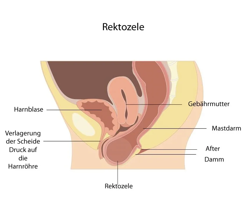 Diagram
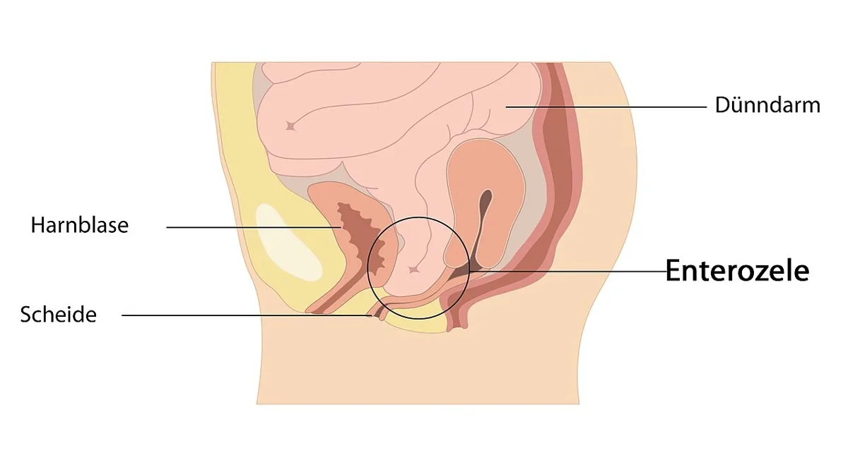 Diagram
Definition
- A rectocele is a form of pelvic organ prolapse in which the rectum herniates through the rectovaginal septum into the posterior vaginal wall.
- It frequently coexists with prolapse of the anterior, apical or posterior compartment, for example an enterocele, in which bowel herniates into the vaginal canal through the pouch of Douglas.
Occurrence & epidemiology
Epidemiology
- Up to two thirds of parous women have some degree of rectocele, most of them without symptoms.
- A rectocele may also develop in about 12% of nulliparous women, attributed to congenital defects.
Aetiopathogenesis
Aetiology and pathogenesis
- Weakening or disruption of the rectovaginal septum allows the anterior rectal wall to herniate into the vagina; the most common defect is a transverse break just above the perineal body.
- Weakening of the levator ani muscles or the surrounding connective tissue also contributes.
- Non-modifiable risk factors are advanced age and genetic susceptibility; further factors are higher parity, vaginal birth, obesity, constipation and chronically raised intra-abdominal pressure.
Clinical features
- Typical are a posterior vaginal bulge that worsens with straining, terminal constipation with obstructed defecation, perineal pressure and vaginal or pelvic discomfort.
- A sense of incomplete bowel emptying is common; some patients insert fingers into the vagina and press on the posterior wall (splinting) to change the rectal angle and defecate.
- In advanced cases the prolapse extends beyond the hymen, and the exposed mucosa may erode, bleed, become infected or ulcerate.
Diagnosis
- Examination includes vaginal and rectal assessment: with the anterior wall retracted, the posterior vaginal wall is observed at rest and on straining, together with a rectovaginal examination.
- In the POP-Q system, the rectocele is assessed using two measurement points on the posterior vaginal wall and then staged.
- Fluoroscopic defecography is usually used for anatomical assessment, and dynamic MR defecography gives more detailed images; ultrasonography, anorectal manometry or colonic transit studies may be added.
- Radiological evidence of rectocele is non-specific, as defecography shows it in up to 93% of healthy, asymptomatic women.
Keep learning in the app
Further reading (selection)
Cross-references
Note: Learning content from the GynFuchs app (flashcards, exam questions, image cases) for medical education – not a treatment recommendation and no substitute for diagnosis or treatment decisions in individual cases. Treatment and management are deliberately not covered on this page.