Cystocele
Exam relevance: in 15 of 197 board exam reports · rank 49
- Specialty
- Gynaecology · Pelvic floor & urogynaecology
- Images
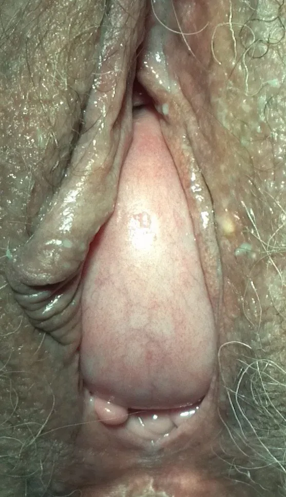
- Colposcopy 1
- Exam relevance
- 15 of 197 reports · rank 49
- In the app
- 1 flashcards · GynFuchs
- Last updated
- 10/2026 · Dr. Pascal Bafteh
Contents
Images (1)
Definition
- Prolapse of the anterior vaginal wall in which the bladder protrudes into the vagina; if the urethra is also involved, it is called a cystourethrocele.
- A cystocele commonly develops when the pubocervical fascia is weakened.
Classification
Two Defects
- Pulsion cystocele: a central defect with attenuated pubocervical fascia.
- Traction cystocele: a lateral defect with avulsion of the fascia from the arcus tendineus fasciae pelvis.
- Characteristic of a pulsion cystocele in the image: the rugae of the vaginal wall are effaced over the bulge.
- Characteristic of a traction cystocele in the image: the rugae are preserved on the bulging wall.
- In the POP-Q system, points Aa and Ba describe the anterior vaginal wall.
- POP-Q stages range from 0 (no prolapse) through I (most distal point more than 1 cm above the hymen), II (between 1 cm above and 1 cm below) and III (more than 1 cm below but at least 2 cm short of total vaginal length) to IV (complete eversion).
- The older Baden-Walker system, based on the level of protrusion, is considered imprecise and only moderately reproducible and is no longer commonly used.
Occurrence & epidemiology
Epidemiology
- Pelvic organ prolapse is common, but its prevalence is difficult to determine; in a study of 8000 women, 8.3% reported symptomatic prolapse.
- Usually several sites are involved; cystocele, enterocele and rectocele are particularly likely to occur together.
Aetiopathogenesis
Aetiology and pathogenesis
- A common risk factor is the number of vaginal births, particularly with a prolonged second stage of labour, instrumental vaginal birth or a large-for-gestational-age newborn.
- Other factors are obesity, increasing age and chronically raised intra-abdominal pressure from constipation, heavy lifting or chronic respiratory disorders; sacral nerve disorders and connective tissue disorders are less common.
Clinical features
- Typical are pelvic or vaginal fullness, pressure and a sensation of organs falling out; the bulge appears particularly on straining or coughing, and mild prolapse may be asymptomatic.
- Insufficient urethral support may cause stress incontinence, whereas marked prolapse can kink the urethra, improve incontinence symptoms and even lead to urinary retention.
- Prolapse may negatively affect body image and be associated with dyspareunia and sexual dysfunction.
Diagnosis
How to Differentiate on the Examination Chair
- Helpful manoeuvre: lifting the lateral sulci with ring forceps — the traction cystocele disappears.
- Examination condition: the assessment is performed during straining and with a moderately full bladder.
- Honest assessment: the clinical classification is uncertain and has high inter-observer variability.
Typical imaging findings
- Clinical finding: On straining the anterior vaginal wall bulges as a soft, smooth hemisphere to the level of the introitus. No cervix is visible on this structure and the posterior wall stays in place.
- The diagnosis is clinical: the posterior vaginal wall is retracted with a speculum blade and the anterior wall is assessed at rest and on straining.
- Prolapse appears more severe when the examination is done upright or standing rather than supine.
- Occult stress incontinence is urine loss on cough testing with the prolapse reduced in women who do not report incontinence.
Keep learning in the app
Further reading (selection)
- MSD Manual Professional: Anterior and Posterior Vaginal Wall Prolapse
- MSD Manual Professional: Overview of Pelvic Organ Prolapse (POP)
- International Urogynecology consultation chapter 2 committee 3: the clinical evaluation of pelvic organ prolapse including investigations into associated morbidity/pelvic floor dysfunction (Int Urogynecol J 2023, PubMed Central)
- DocCheck Flexikon, Zystozele
Cross-references
Note: Learning content from the GynFuchs app (flashcards, exam questions, image cases) for medical education – not a treatment recommendation and no substitute for diagnosis or treatment decisions in individual cases. Treatment and management are deliberately not covered on this page.