Meconium-stained amniotic fluid
Exam relevance: in 1 of 197 board exam reports · rank 171
- Specialty
- Obstetrics · Birth & puerperium
- Images
- Histology & cytology 2
- Exam relevance
- 1 of 197 reports · rank 171
- Last updated
- 10/2026 · Dr. Pascal Bafteh
Contents
Images (2)
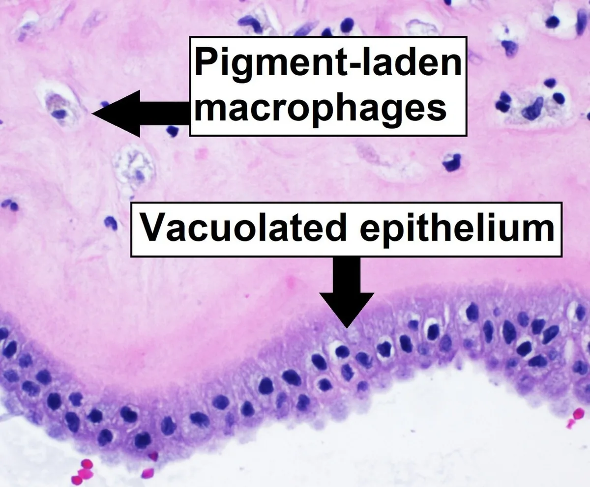 Histology & cytology
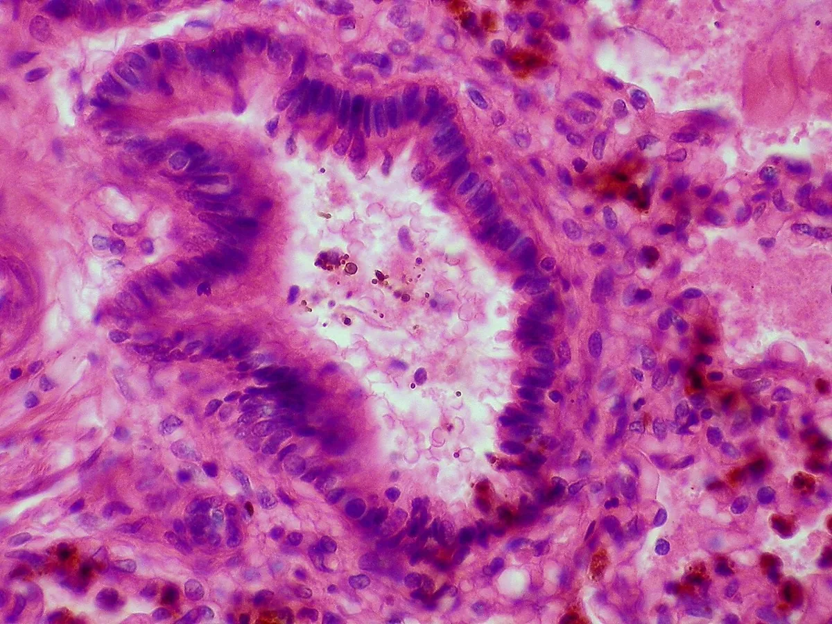 Histology & cytology
Definition
- Meconium-stained amniotic fluid results when the fetus passes meconium, the newborn's first stool, before or during birth.
- Meconium is a black-green, sterile, viscous material consisting of water, desquamated skin and intestinal cells, gastrointestinal secretions, bile, mucus, lanugo, vernix and amniotic fluid.
- Meconium aspiration syndrome is respiratory distress in a newborn with meconium-stained amniotic fluid when the respiratory symptoms cannot be attributed to another cause.
Occurrence & epidemiology
Epidemiology
- Meconium-stained amniotic fluid is found in 4 to 22 per cent of all births and in up to 23 to 52 per cent beyond 42 weeks.
- Only 3 to 12 per cent of infants born through meconium-stained fluid develop meconium aspiration syndrome.
Aetiopathogenesis
Aetiology and risk factors
- Before 34 weeks meconium in the amniotic fluid is uncommon because peristalsis and motilin levels are low and anal sphincter tone is good; meconium aspiration syndrome therefore mainly affects term and post-term newborns.
- In a preterm infant, meconium passage is never considered normal.
- Fetal stress from hypoxia and acidosis, for example with umbilical cord compression or placental insufficiency, or infection leads via a vagal response with increased peristalsis and anal sphincter relaxation to meconium passage.
- Risk factors are placental insufficiency, maternal hypertension, pre-eclampsia, oligohydramnios with cord compression, infections and maternal nicotine and cocaine use.
Clinical features
Clinical features and complications
- Complete bronchial obstruction causes atelectasis, partial obstruction via a ball-valve effect causes air trapping with hyperinflation and air leak such as pneumothorax or pneumomediastinum.
- Persistent pulmonary hypertension of the newborn can ultimately be the more life-threatening complication.
- Clinical signs are tachypnoea, nasal flaring, retractions, cyanosis or desaturation, rales and rhonchi.
Diagnosis
- It is suspected with respiratory distress after birth through meconium-stained fluid; greenish-yellow staining of the cord, nail beds or skin indicates exposure for several hours but is not diagnostic.
- The diagnosis is confirmed by chest radiograph showing hyperinflation, variable areas of atelectasis, patchy or streaky opacities and diaphragmatic flattening.
Keep learning in the app
Cross-references
Note: Learning content from the GynFuchs app (flashcards, exam questions, image cases) for medical education – not a treatment recommendation and no substitute for diagnosis or treatment decisions in individual cases. Treatment and management are deliberately not covered on this page.