Umbilical cord prolapse
Exam relevance: in 4 of 197 board exam reports · rank 111
- Specialty
- Obstetrics · Birth & puerperium
- Images
- Diagram 1
- Exam relevance
- 4 of 197 reports · rank 111
- In the app
- 1 flashcards · GynFuchs
- Last updated
- 10/2026 · Dr. Pascal Bafteh
Contents
Images (1)
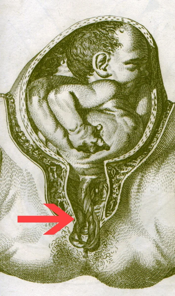 Diagram
Definition
- Cord prolapse means cord is anterior to presenting part after ROM; with intact membranes it is called a cord presentation.
- The danger lies in cord compression with fetal hypoxia.
- The CTG typically shows sudden prolonged or severe variable decelerations.
- In umbilical cord prolapse the cord exits the cervix before the presenting part; it is a rare obstetric emergency with high fetal morbidity and mortality.
Classification
- If the cord lies between the presenting part and the cervix regardless of membrane status, this is cord presentation; descent of the cord through the cervix is essential for prolapse.
- In overt prolapse the cord lies ahead of the presenting part and may protrude from the vagina; in occult prolapse it lies alongside it within the uterus and is often compressed by the shoulder or head.
Occurrence & epidemiology
Epidemiology
- The overall incidence is reported as 0.1–0.6%, higher in non-cephalic presentations, multiple pregnancies and earlier gestational ages; it has fallen markedly over the decades.
Aetiopathogenesis
Risk factors
- They include malpresentation, polyhydramnios, prematurity.
- So for amniotomy: only with a fixed presenting part.
Aetiology and risk factors
- Risk factors are malpresentation, especially in multiparous women, polyhydramnios, multiple pregnancy (especially the second twin), preterm labour and preterm prelabour rupture of membranes; overt prolapse is more common with breech presentation and transverse lie and, in vertex presentation, occurs mainly after rupture of membranes before the head is engaged.
- Up to 50% of cases are iatrogenic, for example from amniotomy, fetal blood sampling or insertion of a cervical balloon.
Clinical features
Clinical features and complications
- Compression of the cord by the descending fetus leads to fetal hypoxia and bradycardia, potentially resulting in death or permanent disability.
- In occult prolapse, a fetal heart rate pattern suggesting cord compression, such as severe bradycardia or severe variable decelerations, is often the only clue.
Diagnosis
- The diagnosis is made when the prolapsed cord is seen or felt in or outside the vagina, together with abnormal fetal heart rate patterns.
- Other causes of sudden fetal bradycardia such as maternal hypotension, placental abruption or uterine rupture are excluded.
Keep learning in the app
Further reading (selection)
Cross-references
Note: Learning content from the GynFuchs app (flashcards, exam questions, image cases) for medical education – not a treatment recommendation and no substitute for diagnosis or treatment decisions in individual cases. Treatment and management are deliberately not covered on this page.