Shoulder dystocia
Exam relevance: in 33 of 197 board exam reports · rank 20
- Specialty
- Obstetrics · Birth & puerperium
- Images
- Mammography/MRI 1 · Clinical 1 · Diagram 1
- Exam relevance
- 33 of 197 reports · rank 20
- In the app
- 2 flashcards · GynFuchs
- Last updated
- 10/2026 · Dr. Pascal Bafteh
Contents
Images (3)
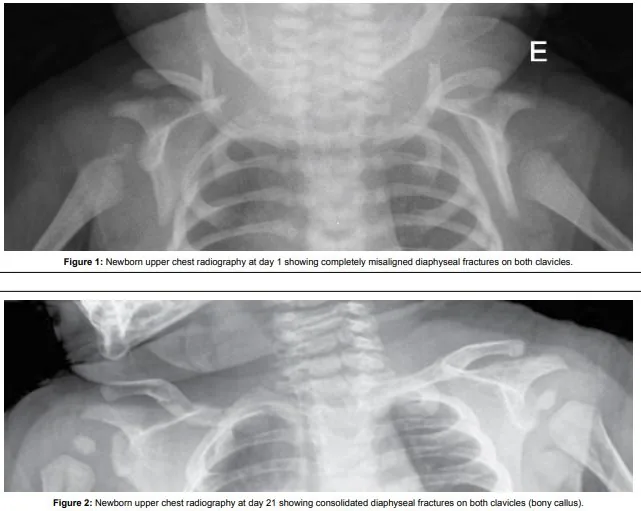 Mammography/MRI
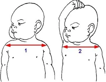 Diagram
Definition
- A distinction is made between high bisacromial and deep transverse arrest; the more dangerous form is the high bisacromial arrest.
- The turtle neck sign describes the retraction of the head against the perineum.
- In shoulder dystocia, after birth of the head the anterior shoulder becomes impacted behind the pubic symphysis, less commonly the posterior shoulder behind the sacral promontory.
- It is characterised by failure of the shoulders to be born with gentle downward traction and the need for additional obstetric manoeuvres; a head-to-shoulder interval of more than 60 seconds has been proposed as an objective criterion.
Occurrence & epidemiology
Figures
- Overall, shoulder dystocia occurs in 0.5–1% of all vaginal births.
- The recurrence risk is at approx. 14%.
Epidemiology
- Shoulder dystocia occurs in about 0.2 to 3% of vaginal births in vertex presentation.
- The incidence rises with birth weight from about 0.3–1% at 2,500 to 4,000 g to 5–7% at 4,000 to 4,500 g; nevertheless, more than half of cases involve infants of normal birth weight.
Aetiopathogenesis
Risk factors
- They include macrosomia, diabetes, obesity, prolonged labour.
- In diabetic women the risk is higher because the trunk grows disproportionately.
Aetiology and risk factors
- The cause is a shoulder (biacromial) diameter that is too large relative to the birth canal.
- Risk factors are fetal macrosomia, maternal obesity, diabetes mellitus, shoulder dystocia in a previous pregnancy, forceps or vacuum birth and very rapid or prolonged labour.
- An early warning sign is a prolonged second stage, particularly in fetuses with risk factors.
Clinical features
More facts from the study questions
- The recurrence risk for shoulder dystocia is stated to be approximately 14 percent.
- In pregnant women with diabetes mellitus, disproportionate growth of the foetal trunk is common.
- This leads to broader shoulders and increases the risk of shoulder dystocia.
- The necessary manoeuvres and the prolonged second stage of labour increase the risk for the mother.
- Typical maternal complications include uterine atony and high-grade perineal or cervical tears.
- Although macrosomia is a significant risk factor, the majority of shoulder dystocias occur unexpectedly.
- Most cases happen in infants with a normal birth weight.
- The aim is to adduct the anterior shoulder and rotate it under the symphysis pubis.
- The Kristeller manoeuvre and traction on the head are strictly contraindicated in shoulder dystocia.
- Both manoeuvres can worsen the shoulder impaction and lead to severe injury of the fetal brachial plexus.
Clinical features and complications
- The born head may retract against the perineum (turtle sign); however, only some cases show this sign.
- Brachial plexus injuries occur in about 7–20% of shoulder dystocias, more often on the right; the most common is upper plexus palsy (Erb, C5–C6), less often lower plexus palsy (Klumpke, C8–T1) or complete palsy; most resolve within 6–12 months, but about 10% of Erb palsies do not.
- Further neonatal consequences are clavicular and humeral fractures and, with prolonged dystocia, fetal compromise up to asphyxia and death.
- Maternal complications are postpartum haemorrhage, perineal tears, anal sphincter injuries, symphysis pubis separation and lateral femoral cutaneous neuropathy.
Diagnosis
- Shoulder dystocia is a clinical diagnosis made only when it occurs: the head has been born but the anterior shoulder does not follow despite gentle traction.
- Fetal weight alone is not a reliable predictor.
Keep learning in the app
Further reading (selection)
Cross-references
Note: Learning content from the GynFuchs app (flashcards, exam questions, image cases) for medical education – not a treatment recommendation and no substitute for diagnosis or treatment decisions in individual cases. Treatment and management are deliberately not covered on this page.