Hydatidiform mole (gestational trophoblastic disease)
Exam relevance: in 5 of 197 board exam reports · rank 101
- Synonyms
- molar pregnancy, hydatidiform mole
- Specialty
- Obstetrics · Early pregnancy
- Images
- Ultrasound 3 · Endoscopy & gross 3
- Exam relevance
- 5 of 197 reports · rank 101
- In the app
- 1 flashcards · GynFuchs
- Last updated
- 10/2026 · Dr. Pascal Bafteh
Contents
Images (6)
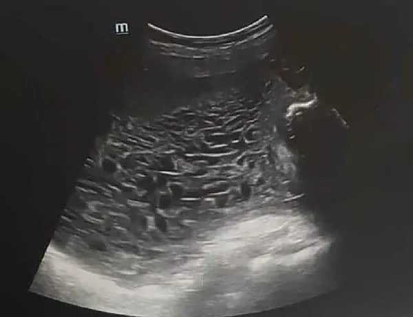 Ultrasound
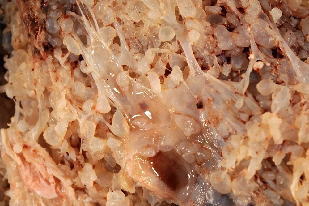 Endoscopy & gross
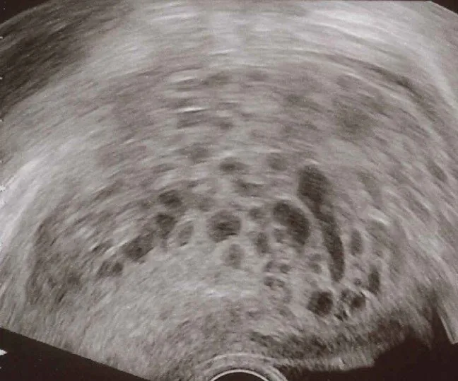 Ultrasound
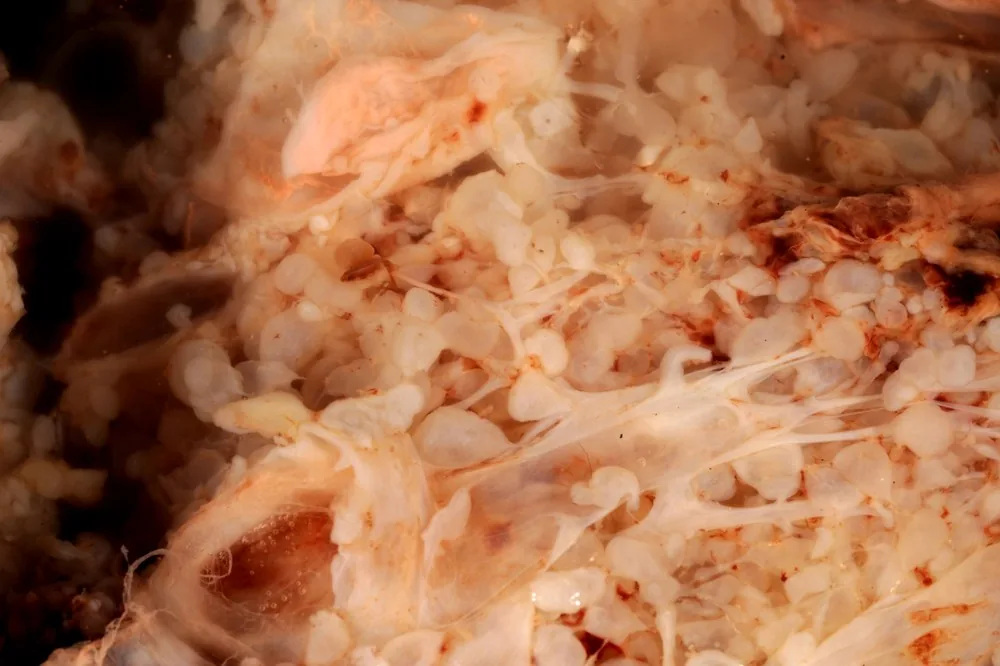 Endoscopy & gross
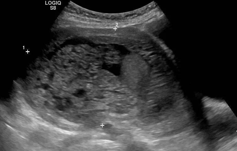 Ultrasound
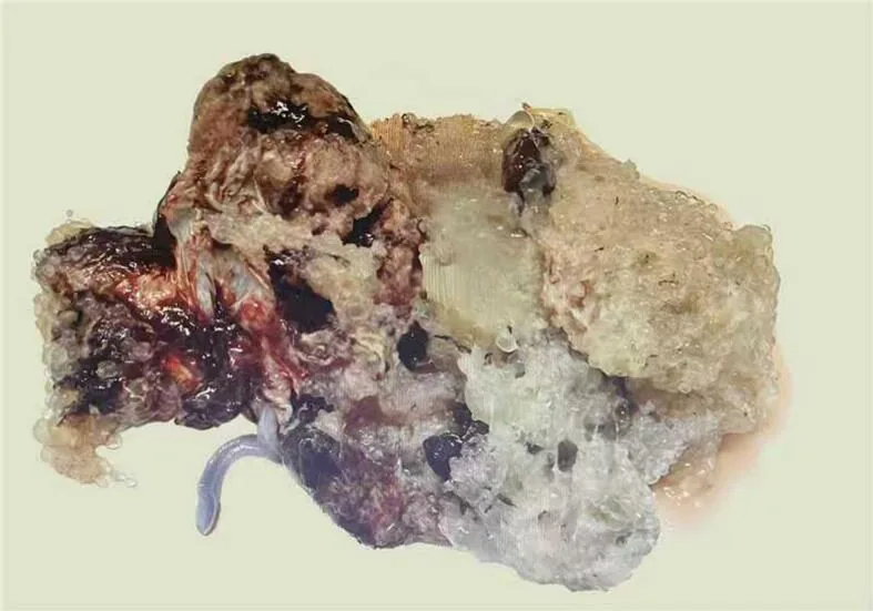 Endoscopy & gross
Classification
Classification and pathogenesis
- A distinction is made between partial and complete hydatidiform moles and gestational trophoblastic neoplasias, which include the invasive mole, choriocarcinoma, placental site trophoblastic tumour, and the epithelioid trophoblastic tumour.
- The complete hydatidiform mole is genetically diploid, purely paternal and typically shows no fetal parts.
- In contrast, the partial mole is triploid with 2 paternal sets and fetal parts may be detectable.
Clinical features
Clinical features and diagnosis
- Clinical signs include bleeding in the first trimester, a uterus that is large for gestational age, hyperemesis, theca lutein cysts, and occasionally hyperthyroidism via hCG cross-reaction.
- Ultrasound classically shows a "snowstorm" or honeycomb pattern in the uterine cavity.
Keep learning in the app
Further reading (selection)
Cross-references
Note: Learning content from the GynFuchs app (flashcards, exam questions, image cases) for medical education – not a treatment recommendation and no substitute for diagnosis or treatment decisions in individual cases. Treatment and management are deliberately not covered on this page.