Gestational trophoblastic neoplasia (choriocarcinoma)
Exam relevance: in 2 of 197 board exam reports · rank 144
- Specialty
- Obstetrics · Early pregnancy
- Images
- Ultrasound 2
- Exam relevance
- 2 of 197 reports · rank 144
- In the app
- 1 flashcards · GynFuchs
- Last updated
- 10/2026 · Dr. Pascal Bafteh
Contents
Images (2)
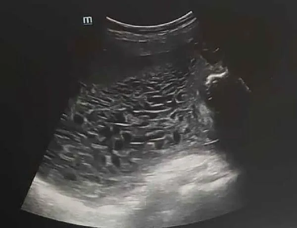 Ultrasound
 Ultrasound
Definition
- Gestational trophoblastic neoplasia (GTN) comprises the malignant, non-molar forms of gestational trophoblastic disease: invasive mole, choriocarcinoma, epithelioid trophoblastic tumour (ETT) and placental site trophoblastic tumour (PSTT).
- Choriocarcinoma is a rare, aggressive neoplasm; the gestational form arises after a hydatidiform mole, a normal pregnancy or a miscarriage.
Classification
Persistent trophoblastic disease
- Criteria for persistent trophoblastic disease include a beta-hCG plateau over three weeks or a/an rise over two weeks.
- Classification uses the anatomical FIGO stages I–IV together with the modified WHO prognostic score; a score of 0–6 is regarded as low risk and 7 or more as high risk.
Occurrence & epidemiology
Epidemiology
- The overall incidence of GTN is about 1 in 40,000 pregnancies.
- After a complete mole about 15% develop an invasive mole and 5% metastatic disease; choriocarcinoma follows 2–3% of hydatidiform moles, more often complete than partial moles.
- Most choriocarcinomas follow a molar pregnancy, miscarriage or ectopic pregnancy; about one quarter are preceded by a full-term pregnancy.
Clinical features
Clinical features and complications
- The leading symptoms of trophoblastic disease are vaginal bleeding and a rapidly enlarging uterus; persistent bleeding after childbirth raises suspicion of choriocarcinoma.
- Choriocarcinoma often first presents with metastases to the lungs, liver or brain, for example with neurological symptoms; it may arise weeks to years after the causative pregnancy.
Histology
- Choriocarcinoma is a malignant tumour of the trophoblastic epithelium: columns and sheets of trophoblastic tissue invade myometrium and blood vessels, with haemorrhage and necrosis.
- Unlike hydatidiform mole and invasive mole, in which villous structures persist, choriocarcinoma lacks villi.
Diagnosis
- Postmolar GTN is usually identified from the hCG course: an hCG plateau over 3 weeks, a rise over 2 weeks (3 values) or hCG still detectable 6 months after the mole; in choriocarcinoma beta-hCG is typically high.
- Staging includes CT of the chest, abdomen and pelvis; pelvic ultrasound or MRI shows the uterine tumour, and metastases outside the lungs usually occur only after lung metastases are established.
Keep learning in the app
Further reading (selection)
Cross-references
Note: Learning content from the GynFuchs app (flashcards, exam questions, image cases) for medical education – not a treatment recommendation and no substitute for diagnosis or treatment decisions in individual cases. Treatment and management are deliberately not covered on this page.