Caesarean scar pregnancy
Exam relevance: in 2 of 197 board exam reports · rank 144
- Specialty
- Obstetrics · Early pregnancy
- Images
- Ultrasound 1
- Exam relevance
- 2 of 197 reports · rank 144
- In the app
- 1 flashcards · GynFuchs
- Last updated
- 10/2026 · Dr. Pascal Bafteh
Contents
Images (1)
Definition
Caesarean scar pregnancy
- Definition: implantation of the gestational sac in the caesarean scar of the lower uterine segment.
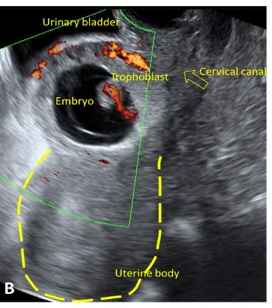
- Ultrasound: an empty cavity and empty cervical canal, the sac in the scar, thin myometrium towards the bladder.
- Danger: massive haemorrhage and uterine rupture as it grows, progression to placenta accreta spectrum.
- Caesarean scar pregnancy is an ectopic pregnancy in which the conceptus implants in the muscle or fibrous tissue of the scar after a previous caesarean section.
Classification
- Following Vial, two types are distinguished: in the first, the pregnancy implanted in the scar develops towards the isthmus and uterine cavity, with a high risk of massive bleeding from the implantation site; in the second, it implants deeply in a damaged scar and leads to uterine rupture and haemorrhage in the first trimester.
Occurrence & epidemiology
Epidemiology
- Its frequency is about 1 in 1,800 to 1 in 2,226 of all pregnancies; after caesarean section it is about 0.15% and accounts for 6.1% of ectopic pregnancies in women with at least one previous caesarean section.
- With rising caesarean section rates and better transvaginal ultrasound it is diagnosed increasingly often.
Clinical features
Clinical features and complications
- Initially it is often asymptomatic; the leading sign is vaginal bleeding without pain.
- Complications include uterine rupture, haemorrhage, coagulation disorders and even maternal death.
Diagnosis
- Typical findings are an empty uterine cavity and cervical canal, a gestational sac or placenta in the caesarean scar, a very thin myometrial layer towards the bladder (about 1–3 to 4.6 mm) and marked vascularity around the scar.
- Colour Doppler shows flow with a peak systolic velocity above 20 cm/s and a pulsatility index below 1; the sliding sign is negative.
- 3D ultrasound and MRI demonstrate the relationship of the sac to the bladder.
Keep learning in the app
Further reading (selection)
Cross-references
Note: Learning content from the GynFuchs app (flashcards, exam questions, image cases) for medical education – not a treatment recommendation and no substitute for diagnosis or treatment decisions in individual cases. Treatment and management are deliberately not covered on this page.