Ectopic pregnancy
Exam relevance: in 32 of 197 board exam reports · rank 23
- Synonyms
- tubal pregnancy, extrauterine pregnancy
- Specialty
- Obstetrics · Early pregnancy
- Images
- Endoscopy & gross 1 · Ultrasound 4
- Exam relevance
- 32 of 197 reports · rank 23
- In the app
- 3 flashcards · GynFuchs
- Last updated
- 10/2026 · Dr. Pascal Bafteh
Contents
Images (5)
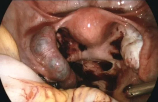 Endoscopy & gross
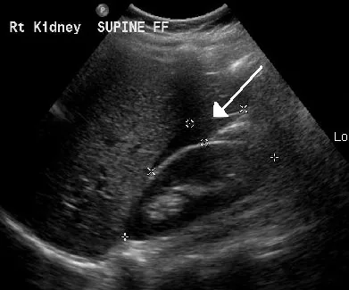 Ultrasound
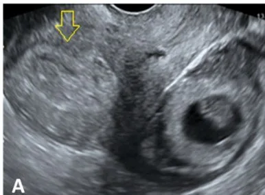 Ultrasound
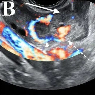 Ultrasound
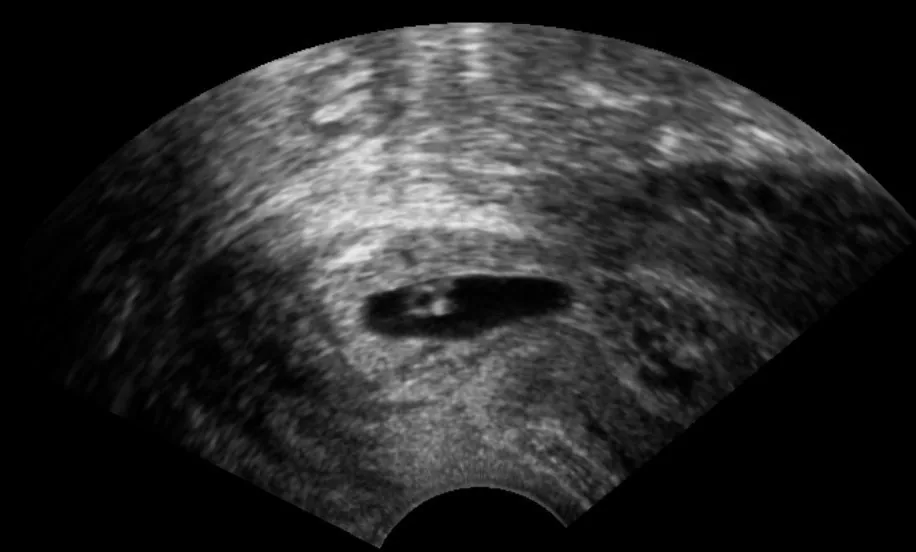 Ultrasound
Definition
Location and frequency
- Ectopic pregnancy (EP) affects approximately 1 to 2% of all pregnancies.
- Over 95% of all EPs are located in the fallopian tube, most commonly in the in the ampullary tubal section.
- The interstitial (cornual) site accounts for about 2–4% of all ectopic pregnancies.
- A rare special form is a heterotopic pregnancy, in which an ectopic pregnancy and an intrauterine pregnancy coexists.
Aetiopathogenesis
Risk factors
- An in-situ intrauterine device (IUD) reduces the absolute number of pregnancies, but if one occurs, the proportion of ectopic implantations is relatively increased.
Clinical features
Presentation and diagnostics
- The cardinal symptoms are secondary amenorrhoea, unilateral lower abdominal pain, and light spotting or bleeding.
- On examination, typical findings include cervical motion tenderness, tenderness in the pouch of Douglas, and often a unilateral, palpable adnexal mass.
- In the event of a rupture, an acute abdomen, haemodynamic instability, and, as a sign of diaphragmatic irritation, unilateral shoulder pain may occur.
- The initial diagnostic steps include checking vital signs, a pregnancy test, determination of blood group and haemoglobin, as well as quantitative hCG and vaginal ultrasound.
More facts from the study questions
- Over 95% of all ectopic pregnancies are located in the fallopian tube.
- Within the tube, the ampullary location is the most frequent.
- A rupture leads to intra-abdominal bleeding (haemoperitoneum).
- The blood can irritate the diaphragm, which can manifest as referred pain in the shoulder (phrenic sign).
- The interstitial, also known as cornual, location is less common.
- It accounts for about 2 to 4% of all ectopic pregnancies.
- An intrauterine device (IUD) very effectively reduces the absolute number of all pregnancies.
- If pregnancy does occur, the proportion of ectopic implantations relative to intrauterine ones is increased.
- A ruptured corpus luteum can also lead to haemoperitoneum and acute lower abdominal pain.
- In an ectopic pregnancy, the uterine cavity remains empty while the endometrium is hormonally stimulated.
Diagnosis
Examination procedure and indirect signs
- An important indirect sign is an empty uterine cavity with a positive pregnancy test and a thickened endometrium.
Classic adnexal findings
- The 'bagel sign' or tubal ring is a round structure with an echogenic ring and an anechoic centre, which is located separate from the ovary.
- When attempting to move the finding with the probe, the bagel sign moves independently of the ovary.
- The 'blob sign' is the most common finding and appears as an inhomogeneous mass next to the ovary.
- The 'ring of fire' on colour Doppler shows strong circular perfusion and is non-specific (also in corpus luteum).
Proof and warning signs
- The only definitive proof of an ectopic pregnancy is the extrauterine sac with embryo/yolk sac.
- This direct proof is only successful in approx. 20% of cases.
- Free fluid in the Pouch of Douglas or Morison's pouch that is echogenic (haemoperitoneum), is a warning sign.
Special forms and inconclusive findings
- In an interstitial pregnancy, the gestational sac is found eccentrically high in the fundus, surrounded by a thin myometrial strip (< 5 mm).
- A caesarean scar pregnancy is present if the gestational sac is located in the niche of the anterior lower uterine segment and there is a very thin myometrium towards the bladder exists.
- In a cervical pregnancy, the gestational sac is located below the internal os with a closed external os and can be distinguished from a cervical abortion by the differentiate by 'sliding sign'.
- An unremarkable ultrasound scan with a positive test does not rule out an ectopic pregnancy; the condition is termed a pregnancy of unknown location.
Beta-hCG trend
- In serum, beta-hCG is detectable approximately 8–9 days after ovulation, whereas urine tests only become reliable after a missed period.
- In a viable early pregnancy, the level rises by at least ≥ 50% in 48 h.
- The peak is reached around the 8th to 10th week, after which the rise flattens.
- An insufficient rise or an overly slow decline suggests a non-viable or ectopic pregnancy, but does not prove its location.
Typical imaging findings
- Ultrasound: Beside the uterus and separate from the ovary lies a round structure with an echogenic ring-like rim and a more hypoechoic centre (tubal ring, bagel sign). The uterine cavity contains no gestational sac; the endometrium is thickened and decidualised.
- Ultrasound: Beside the uterus lies a ring-shaped structure with an echogenic rim. Colour Doppler shows a strong, almost closed vascular ring around it, while the uterine cavity contains no gestational sac.
Keep learning in the app
Further reading (selection)
Cross-references
Note: Learning content from the GynFuchs app (flashcards, exam questions, image cases) for medical education – not a treatment recommendation and no substitute for diagnosis or treatment decisions in individual cases. Treatment and management are deliberately not covered on this page.