Wound complications and niche after caesarean section
Exam relevance: in 6 of 197 board exam reports · rank 93
- Specialty
- Obstetrics · Birth & puerperium
- Images
- Diagram 1
- Exam relevance
- 6 of 197 reports · rank 93
- In the app
- 1 flashcards · GynFuchs
- Last updated
- 10/2026 · Dr. Pascal Bafteh
Contents
Images (1)
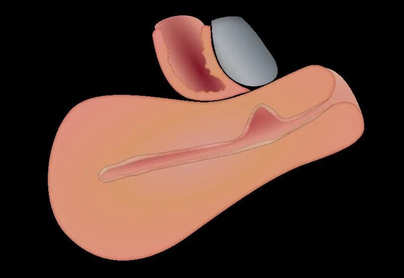 Diagram
Definition
Niche syndrome
- Definition: a defect of the anterior uterine wall at the caesarean scar with thinned myometrium and fluid collection.
- Cardinal symptom: postmenstrual spotting lasting days — blood collects in the niche and drains late.
- Other complaints: chronic pelvic pain, dysmenorrhoea, secondary infertility.
- Work-up: transvaginal ultrasound, plus sonohysterography or hysteroscopy if needed.
- A caesarean scar niche (isthmocele) is an indentation at the site of the caesarean scar with a depth of at least 2 mm.
Classification
- Wound healing disorders after caesarean section comprise superficial infections, wound dehiscence and fluid collections (seroma, haematoma) at the wound site.
- Niches are subclassified as simple, simple with one branch, and complex with more than one branch.
Occurrence & epidemiology
Epidemiology
- Wound healing disorders affect about 3 to 15 per cent of women after caesarean section.
- Niches are frequently detectable on ultrasound, more often with gel or saline contrast than with plain transvaginal sonography.
Aetiopathogenesis
Aetiology and risk factors
- Risk factors for wound complications include young age at childbirth, smoking, obesity, arterial hypertension, diabetes, chorioamnionitis, increased intrapartum blood loss, prolonged rupture of membranes and emergency caesarean section.
Clinical features
Clinical features and complications
- Obese women have an increased risk of wound complications such as haematomas, seromas, abscesses, dehiscence and wound infections.
- Niches are associated with abnormal uterine bleeding, dysmenorrhoea, complications in subsequent pregnancies and possibly subfertility.
Diagnosis
- The niche is examined with transvaginal ultrasound with or without gel or saline contrast, MRI or hysteroscopy; length, depth, residual and adjacent myometrial thickness and width are measured.
Keep learning in the app
Further reading (selection)
Cross-references
Note: Learning content from the GynFuchs app (flashcards, exam questions, image cases) for medical education – not a treatment recommendation and no substitute for diagnosis or treatment decisions in individual cases. Treatment and management are deliberately not covered on this page.