Labour arrest and prolonged labour
Exam relevance: in 15 of 197 board exam reports · rank 49
- Specialty
- Obstetrics · Birth & puerperium
- Images
- Diagram 1
- Exam relevance
- 15 of 197 reports · rank 49
- In the app
- 2 flashcards · GynFuchs
- Last updated
- 10/2026 · Dr. Pascal Bafteh
Contents
Images (1)
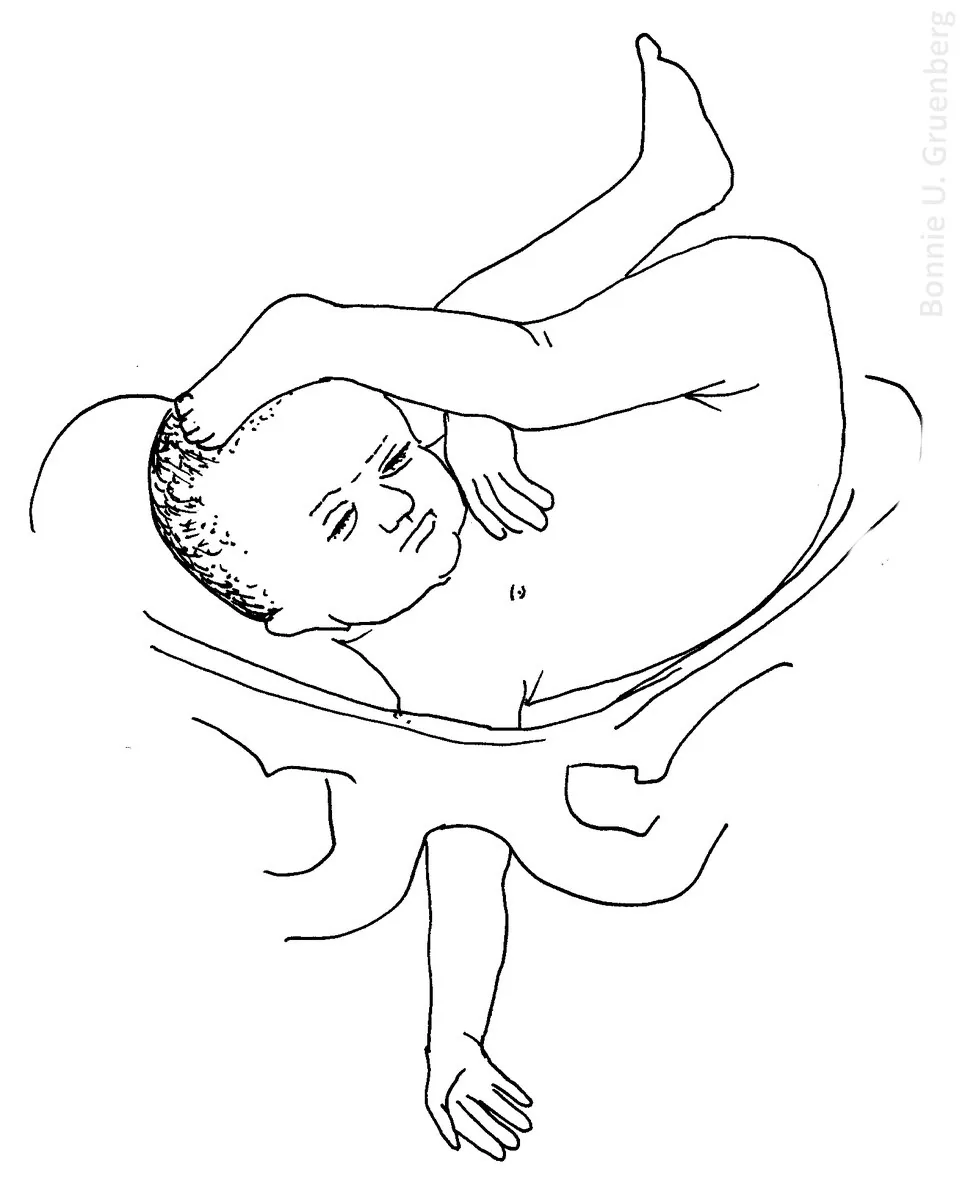 Diagram
Definition
Stages of labour
- The first stage divides into a latent and an active phase (from 5–6 cm).
- The second stage begins at full cervical dilatation.
- A partogram serves graphic documentation of the course of labour.
Arrest of labour
- In the active first stage arrest means at >4 h of no progress despite adequate contractions.
- In the pushing phase the limits are primipara 2 h (epidural 3 h), multipara 1 h (epidural 2 h).
- Arrest in the first stage: no progress in dilatation and station over a defined period despite adequate contractions.
- Arrest in the second stage: no descent despite active pushing over a defined period, longer with an epidural in place than without.
- Before making the diagnosis always check: contraction quality, membrane status, bladder filling, lie and position, analgesia, maternal position.
- Protracted labour is abnormally slow cervical dilatation or fetal descent, labour arrest a complete halt in progress; both can occur in any stage of labour, and uniform definitions are lacking.
- According to ACOG the active first stage begins at 6 cm (5 cm according to WHO); arrest is absent cervical change for 4 hours despite adequate contractions with ruptured membranes, and the latent phase is prolonged beyond 20 hours in nulliparous and 14 hours in multiparous women.
- The second stage is considered prolonged in nulliparous women after more than 3 hours without or more than 4 hours with epidural analgesia, and in multiparous women after more than 2 or 3 hours respectively; the third stage normally lasts no more than 30 minutes.
Aetiopathogenesis
Aetiology and risk factors
- The course of labour depends on the "4 Ps": power (contractions), passage (bony pelvis), passenger (fetus) and presentation.
- Causes are fetopelvic disproportion due to a small pelvis, a large fetus or abnormal presentation, lie or position, and contractions that are too weak or infrequent (hypotonic) or occasionally too strong or frequent (hypertonic).
Clinical features
Uterine inertia
- Primary inertia is present from the onset of labor.
- Causes are exhaustion, uterine overdistension or fear.
Clinical features and complications
- In hypotonic labour, particularly in the active phase, inadequate contractions fail to produce sufficient cervical dilatation and descent; labour becomes protracted.
- Fetal macrosomia is both a cause of protracted labour and a risk factor for shoulder dystocia and severe perineal laceration.
Diagnosis
- The diagnosis is clinical; labour progress is assessed about every 2 to 4 hours, with or without vaginal examination.
- Uterine activity is assessed for strength and frequency by palpation or an intrauterine pressure catheter; clinical or ultrasound estimation of fetal weight helps to identify macrosomia.
Keep learning in the app
Further reading (selection)
Cross-references
Note: Learning content from the GynFuchs app (flashcards, exam questions, image cases) for medical education – not a treatment recommendation and no substitute for diagnosis or treatment decisions in individual cases. Treatment and management are deliberately not covered on this page.