Malposition and malpresentation of the fetal head
Exam relevance: in 7 of 197 board exam reports · rank 88
- Specialty
- Obstetrics · Birth & puerperium
- Images
- Diagram 1
- Exam relevance
- 7 of 197 reports · rank 88
- In the app
- 2 flashcards · GynFuchs
- Last updated
- 10/2026 · Dr. Pascal Bafteh
Contents
Images (1)
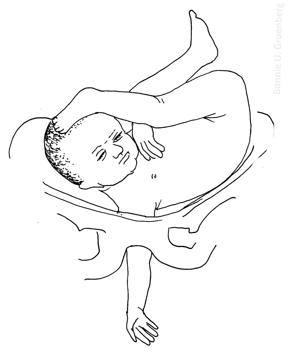 Diagram
Definition
- Malposition and malpresentation of the fetal head are deviations from the normal vertex presentation, in which the flexed head leads with its smallest circumference.
- In face presentation the chin is the presenting part, in brow presentation the area between the anterior fontanelle and the orbital ridges; both result from extension instead of flexion of the neck.
Classification
Malpositions
- In deep transverse arrest of the brim (German "hoher Geradstand") suture sagittal at the pelvic inlet.
- In deep transverse arrest at the outlet the sagittal suture lies transverse at the pelvic outlet.
Malpositions
- High straight position (hoher Geradstand): the head lies in the pelvic inlet in the anteroposterior rather than the transverse diameter — try alternating positions, otherwise caesarean.
- Position describes the relationship of the presenting part to an anatomical axis; in vertex presentation occiput anterior, occiput posterior and occiput transverse positions are distinguished, occiput anterior being normal.
Occurrence & epidemiology
Epidemiology
- Face presentation occurs in about 1 in 600 births; brow presentation is considered the rarest malpresentation (about 1 in 500 to 1 in 4,000 births); the commonest malposition is occiput posterior.
Aetiopathogenesis
Aetiology and risk factors
- Maternal risk factors for face and brow presentation are preterm birth, a contracted or platypelloid pelvis, multiparity and previous caesarean section.
- Fetal risk factors are anencephaly, multiple loops of the umbilical cord around the neck, neck masses, macrosomia and polyhydramnios.
Clinical features
Clinical features and complications
- In occiput posterior position the neck is usually somewhat deflexed, so a larger head diameter passes through the pelvis; labour may arrest in the second stage.
- Brow presentation usually converts spontaneously to vertex or face presentation.
Diagnosis
- The diagnosis is usually made by vaginal examination in the second stage; in face presentation the orbital ridges, nose, malar eminences, mouth and chin are palpable, in brow presentation the anterior fontanelle and face but not the mouth and chin.
- Face presentation may be mistaken for breech; ultrasound shows a reduced angle between occiput and spine or a chin separated from the chest.
Keep learning in the app
Further reading (selection)
Cross-references
Note: Learning content from the GynFuchs app (flashcards, exam questions, image cases) for medical education – not a treatment recommendation and no substitute for diagnosis or treatment decisions in individual cases. Treatment and management are deliberately not covered on this page.