Amniotic fluid embolism
- Specialty
- Obstetrics · Birth & puerperium
- Images
- Histology & cytology 2
- In the app
- 1 flashcards · GynFuchs
- Last updated
- 10/2026 · Dr. Pascal Bafteh
Contents
Images (2)
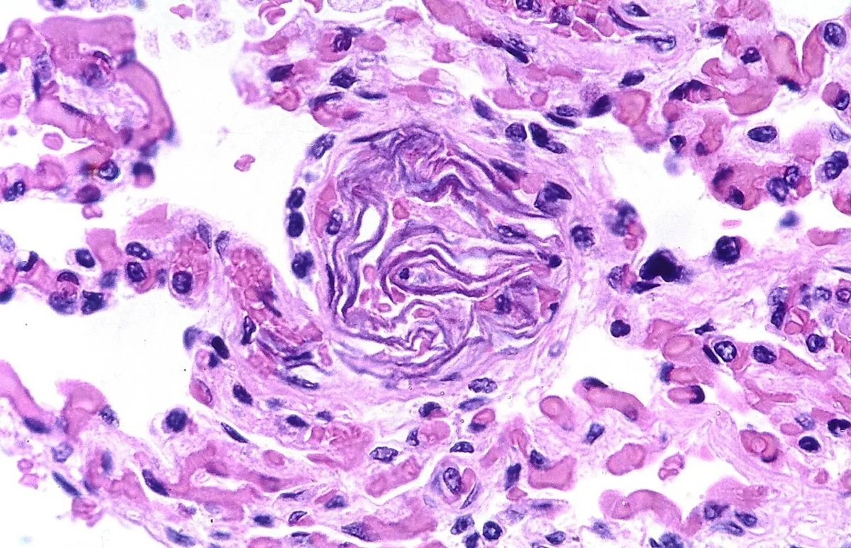 Histology & cytology
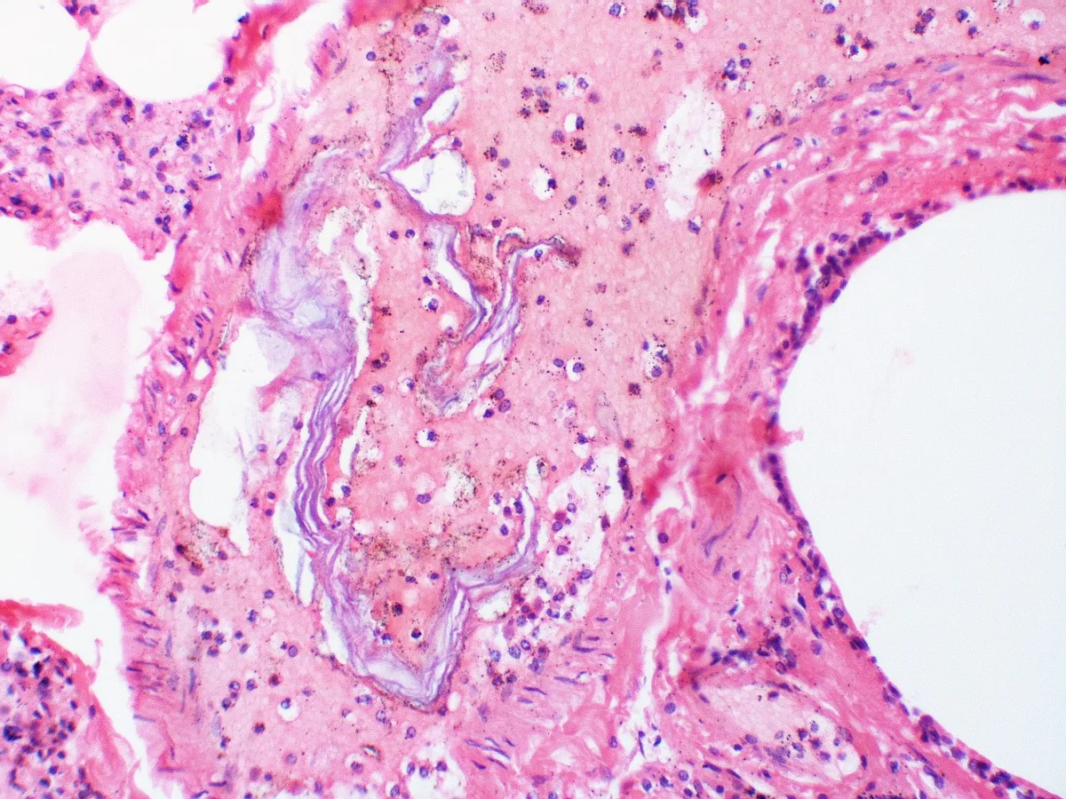 Histology & cytology
Occurrence & epidemiology
Epidemiology
- Incidence estimates vary widely (worldwide 1 in 8,000 to 1 in 80,000 births); for the United States, 2.2 to 7.7 cases per 100,000 births are reported.
- In the United States it is the second leading cause of peripartum maternal death and the primary cause of peripartum cardiac arrest; with caesarean section the incidence in one analysis was about three times that with vaginal birth.
Aetiopathogenesis
Aetiology and risk factors
- Exposure to fetal antigens activates pro-inflammatory mediators and an overwhelming inflammatory response similar to SIRS; this leads to organ damage, especially of the lungs and heart, pulmonary vasoconstriction with right ventricular failure and activation of coagulation up to DIC.
- Risk factors, with inconsistent evidence, include advanced maternal age, multiple pregnancy, polyhydramnios, placenta praevia and accreta, placental abruption, eclampsia, abdominal trauma, cervical lacerations, forceps birth, uterine rupture and caesarean section.
Clinical features
Picture
- Typical is sudden collapse with hypoxia & coagulopathy.
- The sequence includes dyspnoea, cyanosis, hypotension, seizure.
- On the CTG there is often an acute fetal bradycardia.
- The diagnosis is a clinical diagnosis of exclusion.
Clinical features and complications
- It usually occurs during or shortly after labour; the first sign may be sudden cardiac arrest, otherwise dyspnoea, tachycardia, tachypnoea and hypotension develop suddenly, rapidly followed by respiratory failure.
- About one third of patients previously have chills, agitation, a sudden feeling of anxiety, nausea or altered mental status; stroke and seizures are less common.
Diagnosis
- The diagnosis is clinical and one of exclusion: it is suspected when the classic triad develops during or immediately after labour, and made after other causes of haemodynamic instability have been excluded.
Keep learning in the app
Further reading (selection)
Cross-references
Note: Learning content from the GynFuchs app (flashcards, exam questions, image cases) for medical education – not a treatment recommendation and no substitute for diagnosis or treatment decisions in individual cases. Treatment and management are deliberately not covered on this page.