Pityriasis rubra pilaris
Definition & Pathogenesis
- Pityriasis rubra pilaris (PRP) = chronic inflammatory, papulosquamous skin disease, clinically positioned between psoriasis and ichthyosis.
- Pathogenesis: The driving force is the IL-23/Th17 axis with an increase in IL-17/IL-23 — hence the response to IL-17/IL-23 inhibitors.
- Genetic: familial form (5% of cases) — autosomal dominant mutation in the CARD14 gene (Type V), same locus as in familial psoriasis.
Clinical Presentation — Characteristic Triad
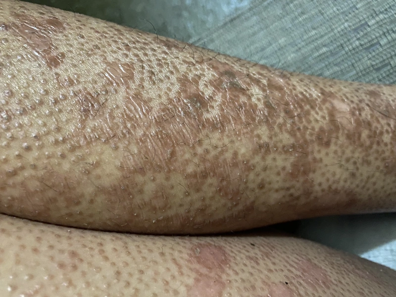
- 1. Hyperkeratotic follicular papules with central keratosis ("Karelle" — grain-like horny plugs).
- 2. Confluent pityriasiform plaques — orange-reddish in color.
- 3. Salmon-colored erythematokeratotic areas with pathognomonic characteristics.
Pathognomonic Findings
- Pathognomonic are nappes claires — i.e., islands of normal skin within the plaques.
- Salmon-colored (orange-red) plaques as a typical hue — together with the nappes claires, the orange-red colour of the (sub-)erythroderma is the key clinical clue.
- Typical are follicular horny plugs with central keratosis.
- Subungual hyperkeratosis similar to psoriasis — but NO pitting nails and NO oil drop phenomena (DD to psoriasis!).
- Palmoplantar keratosis ("PRP keratosis") — orange-yellowish, sharply demarcated, often waxy ("sandal-like" keratoderma).
- Negative Auspitz phenomenon ( in contrast to psoriasis vulgaris negative).
Classification according to Griffiths (Subtypes I–VI)
| Type | Designation | Characteristics | Frequency |
|---|---|---|---|
| I | Classic adult type | acute, generalized, good prognosis, spontaneous remission in 1–3 years | ~ 55 % (most common form!) |
| II | Atypical adult | chronic (decades), atypical, ichthyosis-like, alopecia | ~ 5 % |
| III | Classic juvenile type | acute post-infectious (streptococcal / viral triggers), children 5–10 years, good prognosis | ~ 10 % |
| IV | Circumscribed juvenile form | knees / elbows, localized, preadolescent | ~ 25 % |
| V | Atypical juvenile (familial, CARD14 mutation) | chronic, from birth, finger-like scleroderma-like lesions | < 5 % |
| VI | HIV-associated type | aggressive course, conjunctivitis, difficult to treat | rare |
Histology
- Histologically typical is the checkerboard pattern of alternating ortho- and parakeratosis.
- Acanthosis, follicular plugging, focal vascular dilatation.
- Negative: Munro microabscesses (DD: positive in psoriasis).
Diagnostics
- Clinical presentation primarily (triad + nappes claires + salmon-colored plaques).
- Punch biopsy for histological confirmation.
- In case of suspected Type III (children): ASO titer, throat swab (streptococci), virology.
- In case of suspected Type VI: HIV serology obligatory.
- In familial / atypical form: CARD14 sequencing.
Differential diagnoses
- Psoriasis vulgaris (plaque psoriasis)
- Seborrhoeic dermatitis
- Mycosis fungoides
- Lichen planus
- Ichthyosis
- Atopic dermatitis (atopic eczema)
Practise Pityriasis rubra pilaris in the app
Flashcards with spaced repetition, exam questions and spot-the-diagnosis on this topic – in DermaFuchs, free of charge.
In the DermaFuchs app: 3 flashcards · 6 clinical images
Open in browser Download on the App StoreSources (selection)
More inflammatory skin diseases
- Cheilitis granulomatosa (Miescher)
- Erythema annulare centrifugum (EAC)
- Erythema nodosum
- Graft-versus-host disease (GvHD)
- Granuloma annulare
- Cutaneous sarcoidosis
- Lichen planus
- Lichen sclerosus
Note: This page is intended for medical education and does not replace diagnosis or treatment decisions in individual cases. Treatment and follow-up content is available in the app.