Ventriculomegaly and hydrocephalus
Exam relevance: in 2 of 197 board exam reports · rank 144
- Specialty
- Obstetrics · Fetus & prenatal medicine
- Images
- Ultrasound 1
- Exam relevance
- 2 of 197 reports · rank 144
- Last updated
- 10/2026 · Dr. Pascal Bafteh
Contents
Images (1)
Definition
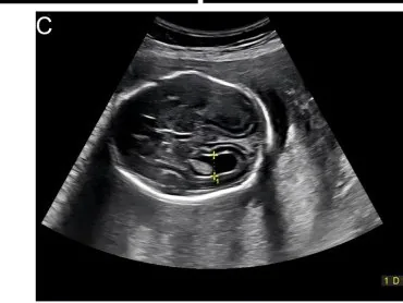
- Fetal ventriculomegaly is a prenatally detected enlargement of the lateral ventricles with a diameter of 10 mm or more in the axial transventricular plane.
- Hydrocephalus denotes a symptomatic accumulation of CSF in the cerebral ventricles due to obstructed CSF flow or impaired absorption.
Classification
- By ventricular diameter, mild (10 to 12 mm), moderate (13 to 15 mm) and severe forms (16 mm or more) are distinguished.
- It is considered isolated when no further structural or genetic abnormalities are identified on ultrasound.
Occurrence & epidemiology
Epidemiology
- It occurs in about 2 per 1,000 live births and is one of the most common fetal anomalies detected in the second trimester.
Aetiopathogenesis
Aetiology
- Causes are parenchymal loss, obstruction and overproduction of CSF; the most common structural causes are aqueductal stenosis, Chiari II malformation, dysgenesis of the corpus callosum and posterior fossa anomalies.
- Chromosomal abnormalities are found in 2 to 12%, most commonly trisomy 21; intrauterine infections such as toxoplasmosis, rubella, CMV and herpes simplex in about 5%.
Clinical features
- Neurodevelopment depends mainly on severity and associated anomalies; severe forms usually have obstructive causes and are associated with hydrocephalus.
Diagnosis
- The normal ventricular diameter is 4.5 to 7.6 mm between 15 and 40 weeks; the atrium remains stable during the second and third trimesters.
- Fetal MRI detects additional abnormalities in about 50% of cases; genetic testing uses karyotyping and chromosomal microarray.
Keep learning in the app
Further reading (selection)
Cross-references
Note: Learning content from the GynFuchs app (flashcards, exam questions, image cases) for medical education – not a treatment recommendation and no substitute for diagnosis or treatment decisions in individual cases. Treatment and management are deliberately not covered on this page.