Trisomy 21 (prenatal diagnosis)
Exam relevance: in 9 of 197 board exam reports · rank 75
- Synonyms
- Down syndrome, trisomy 21
- Specialty
- Obstetrics · Fetus & prenatal medicine
- Images
- Ultrasound 3 · Clinical 1
- Exam relevance
- 9 of 197 reports · rank 75
- In the app
- 1 flashcards · GynFuchs
- Last updated
- 10/2026 · Dr. Pascal Bafteh
Contents
Images (4)
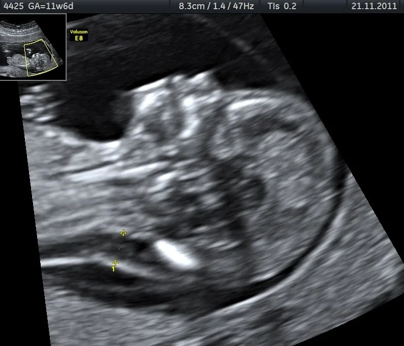 Ultrasound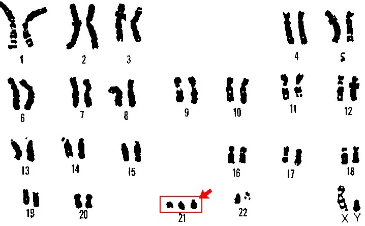
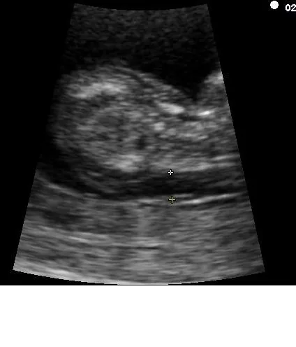 Ultrasound
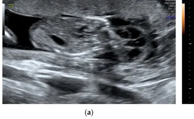 Ultrasound
Definition
- Chromosomal aberration with an extra copy of chromosome 21; it is the most common viable autosomal trisomy and the leading genetic cause of intellectual disability.
Classification
- About 95% result from meiotic non-disjunction, usually of maternal origin, and about 4% from translocations (most commonly t(14;21)); mosaicism due to non-disjunction during embryonic development is less common.
Occurrence & epidemiology
Epidemiology
- In the United States the incidence is about 1 in 700 live births; the risk rises with maternal age from about 1 in 1,476 at 20 through 1 in 352 at 35 to 1 in 85 at 40.
- Because most births occur to younger women, only about 20% of children with Down syndrome are born to mothers over 35.
Clinical features
Prenatal signs and clinical features
- Typical features are intellectual disability, muscular hypotonia, characteristic craniofacial features and various malformations.
- About 50% of neonates have congenital heart disease, most often ventricular septal defect and atrioventricular septal defect; about 6% have gastrointestinal anomalies, particularly duodenal atresia.
Diagnosis
Biochemical markers
- A typical pattern for trisomy 21 is low PAPP-A and high free beta-hCG.
- In trisomy 18 or 13, however, both values are low, which is a clear difference from the pattern in trisomy 21.
- The risk calculation is improved by additional markers such as the nasal bone, ductus venosus and tricuspid regurgitation.
- First-trimester combined screening measures maternal serum beta-hCG and PAPP-A together with nuchal translucency; in trisomy 21, beta-hCG is typically raised, PAPP-A low and nuchal translucency enlarged, although no nuchal translucency threshold is diagnostic.
- Cell-free DNA analysis in maternal plasma is possible from 10 weeks and has higher detection rates than older methods but remains a screening test; the diagnosis is confirmed by karyotyping, prenatally for example after chorionic villus sampling.
Keep learning in the app
Further reading (selection)
Cross-references
Note: Learning content from the GynFuchs app (flashcards, exam questions, image cases) for medical education – not a treatment recommendation and no substitute for diagnosis or treatment decisions in individual cases. Treatment and management are deliberately not covered on this page.