Toxoplasmosis in pregnancy
Exam relevance: in 4 of 197 board exam reports · rank 111
- Synonyms
- toxoplasmosis
- Specialty
- Obstetrics · Pregnancy disorders
- Images
- Clinical 1
- Exam relevance
- 4 of 197 reports · rank 111
- In the app
- 1 flashcards · GynFuchs
- Last updated
- 10/2026 · Dr. Pascal Bafteh
Contents
Images (1)
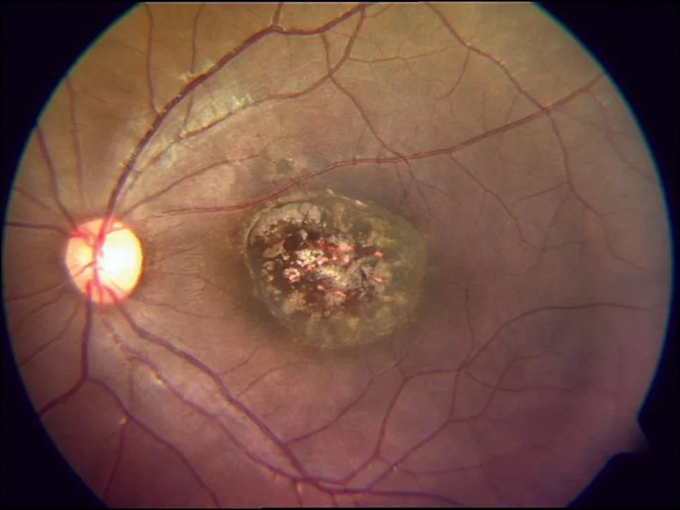
Definition
- Prenatal toxoplasmosis occurs when the mother acquires a first infection with Toxoplasma gondii during pregnancy and the parasite crosses the placenta to the unborn child.
Aetiopathogenesis
Pathogen and Transmission (Logbook 087)
- The pathogen: Toxoplasma gondii.
- Most common route of human infection: ingesting raw meat with tissue cysts.
- Second route: oocysts from cat faeces (soil, vegetables).
- Seroprevalence in Germany: approx. 1/3 to 1/2 of women are immune.
- What is dangerous: only the primary infection during pregnancy.
Aetiology and pathogenesis
- Sources of infection are tissue cysts in raw or undercooked meat and oocysts from cat faeces, for example via contaminated soil during gardening.
- If infection occurred before pregnancy, the fetus is usually protected by maternal antibodies.
- As pregnancy advances, the transmission rate rises from about 15% in the first to about 60% in the last trimester, while the severity of disease in the child decreases.
Clinical features
What to Look for in the Child
- The classic triad of congenital toxoplasmosis: chorioretinitis, hydrocephalus, calcifications.
- Findings on ultrasound may include: ventriculomegaly, calcifications, ascites.
- The most common manifestation overall: silent infection, late chorioretinitis.
Clinical features and complications
- In immunocompetent people, 80–90% do not notice the infection; otherwise a flu-like illness with fever and lymphadenitis, mainly in the head and neck region, occurs.
- Infection in the first third of pregnancy can severely damage the embryo or cause miscarriage.
- The classic triad is retinochoroiditis, hydrocephalus and intracranial calcifications; retinochoroiditis is the most frequent and may appear in initially normal children only after months or years.
Diagnosis
- High IgG avidity excludes infection within the preceding 3–4 months, whereas low avidity does not prove recent infection.
- Prenatally, PCR of amniotic fluid provides evidence of infection of the child; a positive result proves it, a negative one does not reliably exclude it.
Keep learning in the app
Further reading (selection)
Cross-references
Note: Learning content from the GynFuchs app (flashcards, exam questions, image cases) for medical education – not a treatment recommendation and no substitute for diagnosis or treatment decisions in individual cases. Treatment and management are deliberately not covered on this page.