Postmenopausal osteoporosis
Exam relevance: in 4 of 197 board exam reports · rank 111
- Synonyms
- osteoporosis, bone loss
- Specialty
- Gynaecology · Endocrinology & cycle
- Images
- Mammography/MRI 1 · Diagram 1
- Exam relevance
- 4 of 197 reports · rank 111
- Last updated
- 10/2026 · Dr. Pascal Bafteh
Contents
Images (2)
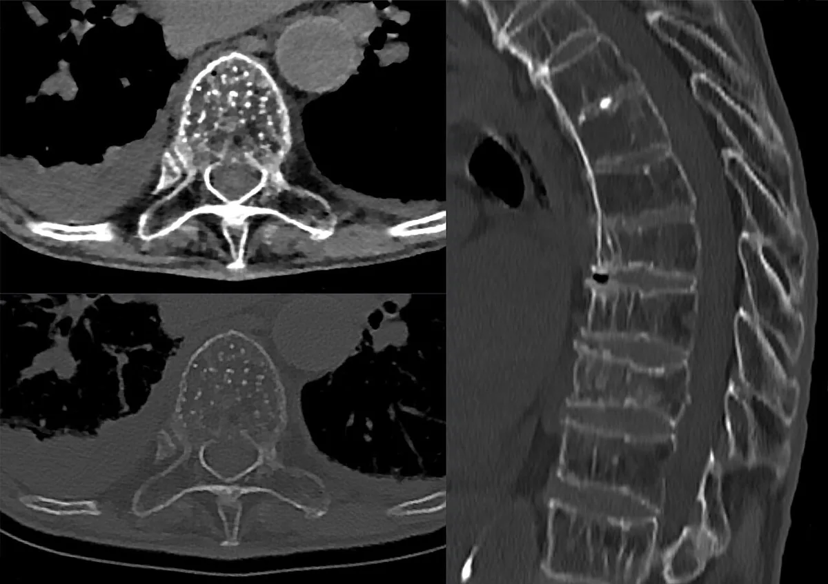 Mammography/MRI
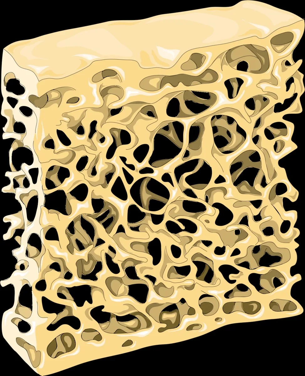 Diagram
Definition
- Osteoporosis is a systemic skeletal disease with low bone mass and distorted microarchitecture, leading to increased fracture susceptibility; in the postmenopausal form, oestrogen deficiency predominates.
- According to the WHO, osteoporosis is present at a DXA T-score of −2.5 SD or lower; severe (established) osteoporosis additionally requires at least one fragility fracture.
Classification
- Primary postmenopausal (type I) osteoporosis is due to oestrogen deficiency; senile (type II) osteoporosis is mainly age-related and affects women about twice as often as men.
- Secondary osteoporosis due to other underlying diseases or substances accounts for up to 30% of osteoporosis in postmenopausal women.
Occurrence & epidemiology
Epidemiology
- Most cases of osteoporosis occur in postmenopausal women and older men; peak bone mass is reached around the age of 30.
Aetiopathogenesis
Aetiology and pathogenesis
- With menopause, bone loss accelerates to about 2% per year for about 10 years; trabecular bone is lost faster than cortical bone because of its higher turnover.
- Risk factors include older age, a positive family history especially of parental hip fracture, low body mass index, immobilisation, smoking, alcohol and insufficient calcium or vitamin D intake.
Clinical features
- The disease is asymptomatic until a fracture occurs; vertebral compression fractures are the most common osteoporotic fractures, but about two-thirds of them remain asymptomatic.
- Typical fracture sites are the thoracic and lumbar spine, femoral neck and distal radius; multiple vertebral fractures lead to height loss and kyphosis.
Diagnosis
- Bone mineral density is measured by DXA, usually at the spine and hip; if these sites are unsuitable, the distal radius (1/3 radius) is used.
- Regardless of bone mineral density, osteoporosis can also be diagnosed on the basis of a fragility fracture, particularly of the spine, hip, humerus or distal radius.
- Secondary causes are sought with calcium, phosphate, 25-hydroxyvitamin D, liver tests including alkaline phosphatase, parathyroid hormone and 24-hour urine calcium and creatinine, supplemented by TSH and blood count depending on the clinical picture.
Keep learning in the app
Further reading (selection)
Cross-references
Note: Learning content from the GynFuchs app (flashcards, exam questions, image cases) for medical education – not a treatment recommendation and no substitute for diagnosis or treatment decisions in individual cases. Treatment and management are deliberately not covered on this page.