Fetal growth restriction (FGR)
Exam relevance: in 33 of 197 board exam reports · rank 20
- Synonyms
- IUGR, intrauterine growth restriction, small for gestational age
- Specialty
- Obstetrics · Pregnancy disorders
- Images
- Ultrasound 6 · Clinical 2
- Exam relevance
- 33 of 197 reports · rank 20
- In the app
- 4 flashcards · GynFuchs
- Last updated
- 10/2026 · Dr. Pascal Bafteh
Contents
Images (8)
 Ultrasound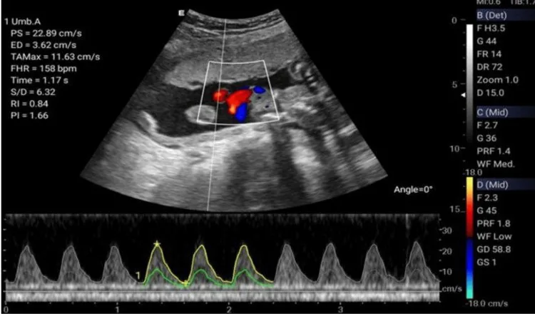
 Ultrasound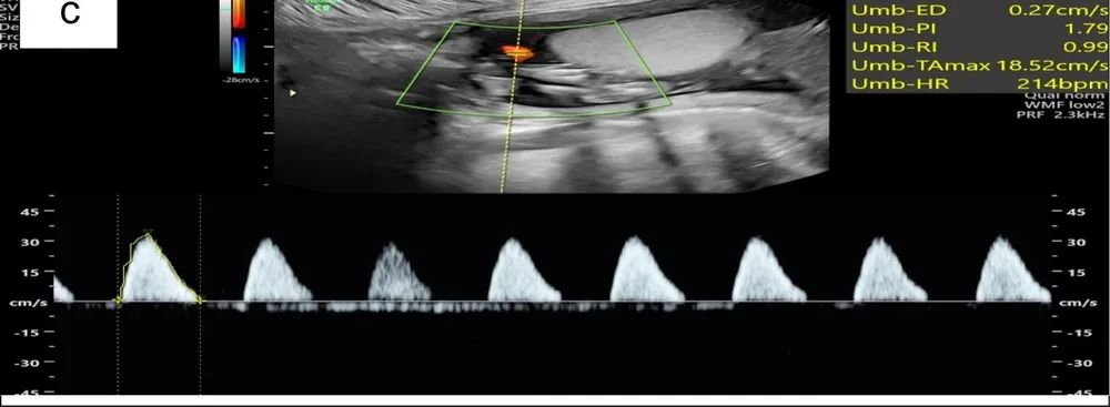
 Ultrasound
 Ultrasound
 Ultrasound
 Ultrasound
Definition
Basic definitions
- A foetus is considered SGA ("small for gestational age") if the estimated foetal weight or abdominal circumference is below the 10th percentile. Some of these babies are constitutionally small and healthy.
- The distinction is clinically relevant because FGR is associated with increased perinatal morbidity and mortality, whereas SGA babies do not have this risk.
Classification
Early versus late onset
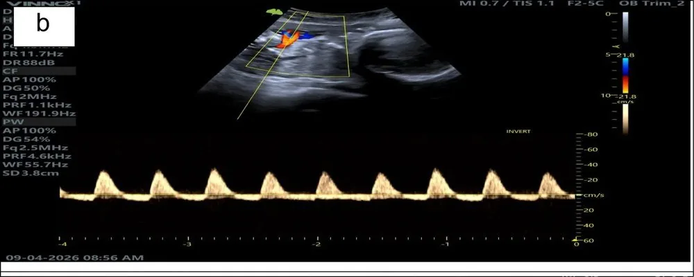
- Early-onset FGR is diagnosed before 32+0 weeks. One criterion is an absent or reversed end-diastolic flow in the umbilical artery.
- Late-onset FGR (from 32+0 weeks) is present with values below the 3rd percentile or if two of three criteria are met: below the 10th percentile, a low cerebroplacental ratio (CPR), or growth faltering >2 quartiles.
- In the early-onset form (before 32+0 weeks), for values below the 10th percentile, a pulsatility index of the umbilical artery or the uterine arteries above the 95th percentile is also sufficient for diagnosis.
Aetiopathogenesis
Causes and morphology
- Symmetrical growth restriction, where all body parts are equally affected, suggests an early cause (genetic, infectious).
- The asymmetrical form with a relatively spared head circumference ("brain sparing") is typical of a late placental cause.
Most common groups of causes
- The most common cause is placental insufficiency, which is based on impaired trophoblast invasion and thus has the same root as pre-eclampsia.
- Among the foetal causes, chromosomal disorders are relevant, especially trisomy 13/18, triploidy.
Clinical features
More facts from the study questions
- A fetus is considered SGA if the estimated weight or abdominal circumference is below the 10th percentile.
- Some of these children are constitutionally small and healthy and have no increased perinatal risk.
- Fetal growth restriction (FGR) is present with an estimated weight or abdominal circumference below the 3rd percentile, even without further criteria.
- Alternatively, the diagnosis can be made with values below the 10th percentile in combination with abnormal Dopplers.
- In early-onset FGR (before 32+0 weeks), a pulsatility index of the umbilical artery or uterine arteries above the 95th percentile is a diagnostic criterion for values below the 10th percentile.
- The reference to the 90th percentile is therefore incorrect.
- The asymmetrical form with a relatively spared head circumference ('brain sparing') is characteristic of a placental cause.
- This form of growth disorder typically manifests at a later stage in the pregnancy.
- The assessment of fetal growth using percentile charts requires the most accurate determination of gestational age possible.
- Without correct dating, the classification of the estimated weight is not reliable.
Diagnosis
Clinical diagnosis of FGR
- The short clinical definition of FGR is an estimated foetal weight or abdominal circumference below the 10th percentile in combination with pathological Doppler findings.
- Alternatively, FGR is also present with an estimated foetal weight or abdominal circumference below the 3rd percentile without other criteria.
- A prerequisite for any percentile specification is correct dating of the pregnancy.
History and basic diagnostics
- In addition to CTG and blood pressure measurement, the initial work-up includes pre-eclampsia labs and a targeted history for risk factors (IUGR, PE, IUFD).
- In the lab, besides kidney and liver function tests, determining the sFlt-1/PlGF ratio is useful for assessing placental function.
Stages of Deterioration
- The first stage of pathology is a pulsatility index above the 95th percentile.
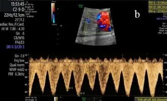
- This is followed by reduced, then absent (AEDF), and finally reversed end-diastolic flow (REDF).
- Absent or reversed end-diastolic flow (ARED flow) means that a large part of the placental vascular bed has failed, meaning the time to fetal acidosis is short.
- With reversed flow, the time to fetal acidosis is shorter than with absent flow.
Measurement and the phenomenon of brain sparing
- The middle cerebral artery (MCA) is insonated in the axial plane near the circle of Willis, where an angle close to zero degrees and no pressure on the fetal head is crucial, as pressure distorts the values.
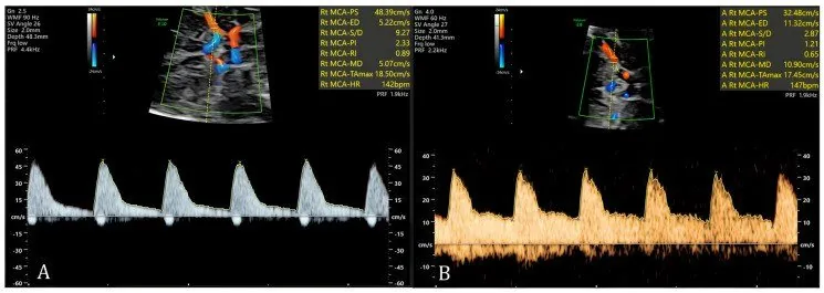
- Brain sparing, also known as centralisation, is a compensatory mechanism in fetal hypoxia where the cerebral vessels dilate. This causes the pulsatility index (PI) of the MCA to falls below the 5th percentile.
- Such a finding is a non-reassuring finding.
Cerebroplacental ratio (CPR)
- The cerebroplacental ratio (CPR) is calculated from the MCA PI divided by umbilical artery PI.
- Values below 1.0 or < 5th percentile are considered pathological.
- The CPR is considered a particularly sensitive marker, especially in late-onset FGR and in post-term pregnancy.
Typical imaging findings
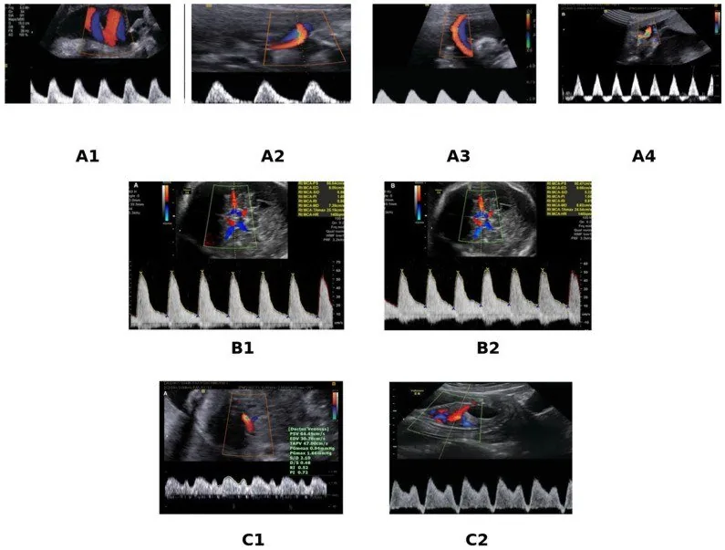
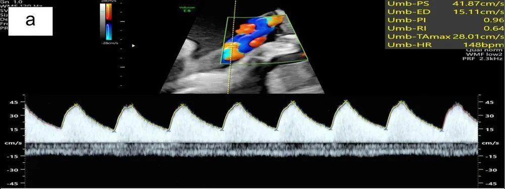
- Ultrasound: The umbilical artery spectrum shows systolic peaks but falls to the baseline in diastole — no forward flow is detectable between heartbeats. The resistance index is correspondingly close to 1.0.
- Ultrasound: The waveform shows tall systolic peaks; in diastole the signal does not merely reach the baseline but crosses it and continues on the opposite side.
- Ultrasound: In the right-hand waveform end-diastolic flow is clearly higher than on the left: the troughs between the peaks stay well above the baseline and the peak-to-trough ratio is strikingly flat. On the left, flow in diastole drops almost to the baseline.
Keep learning in the app
Further reading (selection)
Cross-references
Note: Learning content from the GynFuchs app (flashcards, exam questions, image cases) for medical education – not a treatment recommendation and no substitute for diagnosis or treatment decisions in individual cases. Treatment and management are deliberately not covered on this page.