Systemic mastocytosis
- Synonyms
- mast cell disease, urticaria pigmentosa, cutaneous mastocytosis, mast cell activation syndrome, MCAS, KIT D816V
- Specialty
- Internal medicine · Allergology
- Images
- Clinical 1 · Histology 1
- Last updated
- 10/2026 · Dr. Pascal Bafteh
Contents
Images (2)
 Histology
Definition
Mastocytosis is a group of disorders with clonal proliferation of mast cells and infiltration of the skin, other organs or both. Systemic mastocytosis mostly occurs in adults and is characterised by multifocal mast cell infiltrates in the bone marrow, often with involvement of the skin, lymph nodes, liver, spleen or gastrointestinal tract. Purely cutaneous mastocytosis, in contrast, mainly affects children.
Classification
- Cutaneous mastocytosis: maculopapular cutaneous mastocytosis (formerly urticaria pigmentosa, monomorphic or polymorphic variant), diffuse cutaneous mastocytosis and solitary mastocytoma (1 to 5 cm). In children it rarely progresses to a systemic form, whereas in adults this is possible.
- Indolent systemic mastocytosis: without organ dysfunction.
- Smouldering systemic mastocytosis: high mast cell burden with limited impact on organ function.
- Aggressive systemic mastocytosis: with impaired organ function.
- Systemic mastocytosis with an associated hematological neoplasm: e.g. myeloproliferative neoplasm, myelodysplastic syndrome or lymphoma.
- Mast cell leukemia: more than 20% mast cells in the bone marrow, without skin lesions, with multiorgan failure.
- To be distinguished: mast cell activation syndrome (MCAS), with inappropriate mast cell activation usually without clonal proliferation or organ infiltration.
Aetiopathogenesis
The cause is usually an activating mutation in the KIT gene, which encodes the stem cell factor receptor c-KIT on mast cells; the most common mutation is D816V. It is present in about 80 to 95% of adults and about 30% of children with systemic mastocytosis. Autophosphorylation of the receptor leads to uncontrolled clonal mast cell proliferation.
Symptoms arise mainly from released mediators such as histamine, prostaglandins, leukotrienes and cytokines; extensive organ infiltration can additionally impair organ function. Triggers of mast cell activation include touch and friction, exercise, alcohol, NSAIDs, opioids, insect stings and certain foods.
Clinical features
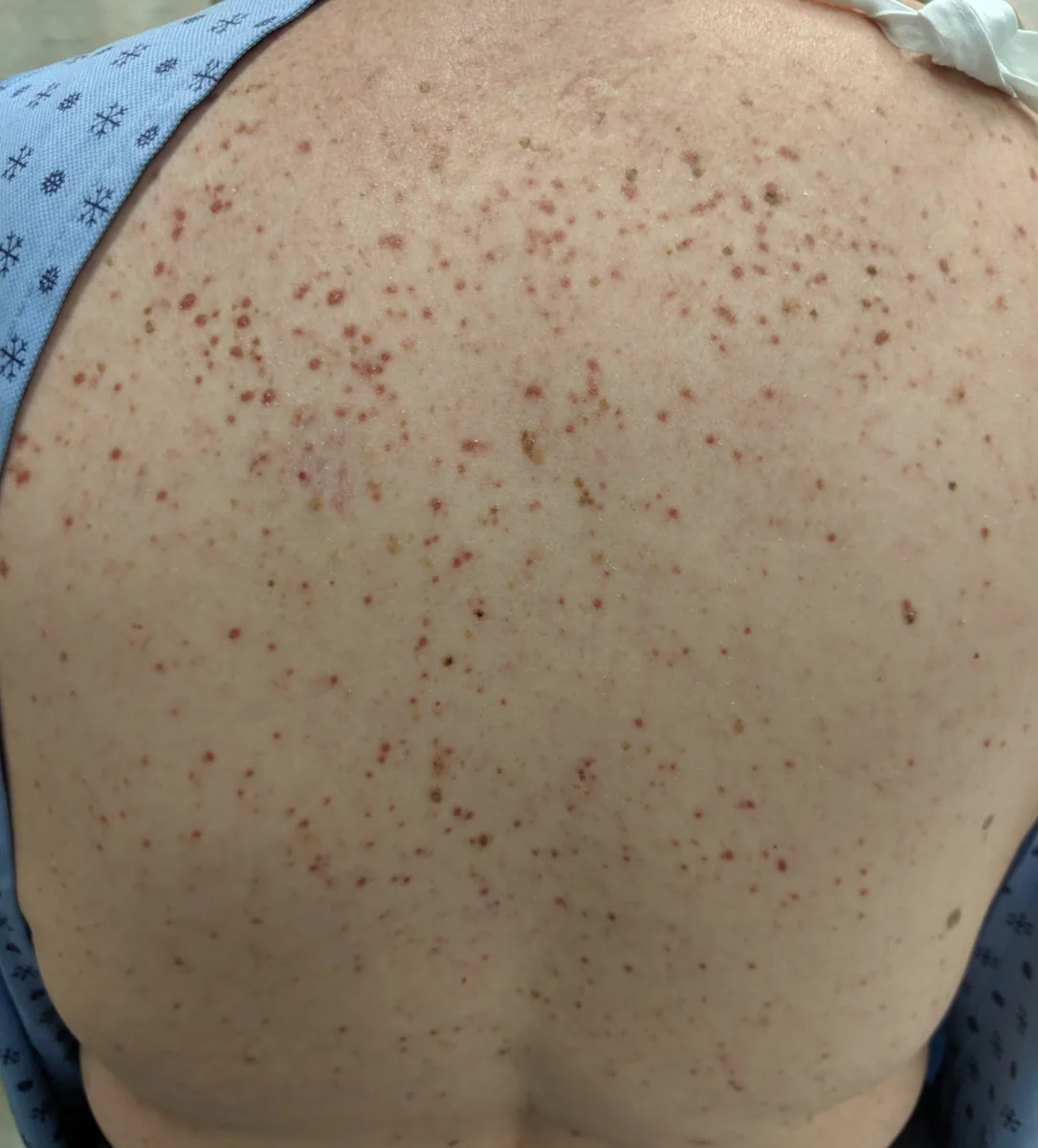
- Skin: salmon-colored to brownish, often itchy maculopapules; rubbing or stroking a lesion causes a wheal and redness around it (Darier sign), unlike dermatographism, which involves normal skin.
- Mediator symptoms: most commonly flushing; anaphylactic reactions with syncope and shock are particularly dangerous.
- Gastrointestinal tract: epigastric pain due to peptic ulcers (histamine stimulates gastric acid secretion), nausea, vomiting and chronic diarrhea.
- Other symptoms: arthralgia, bone pain and neuropsychiatric changes such as irritability, depression and mood swings.
- Organ infiltration: liver and spleen involvement with portal hypertension and ascites; in advanced forms cytopenias due to bone marrow failure.
Histology
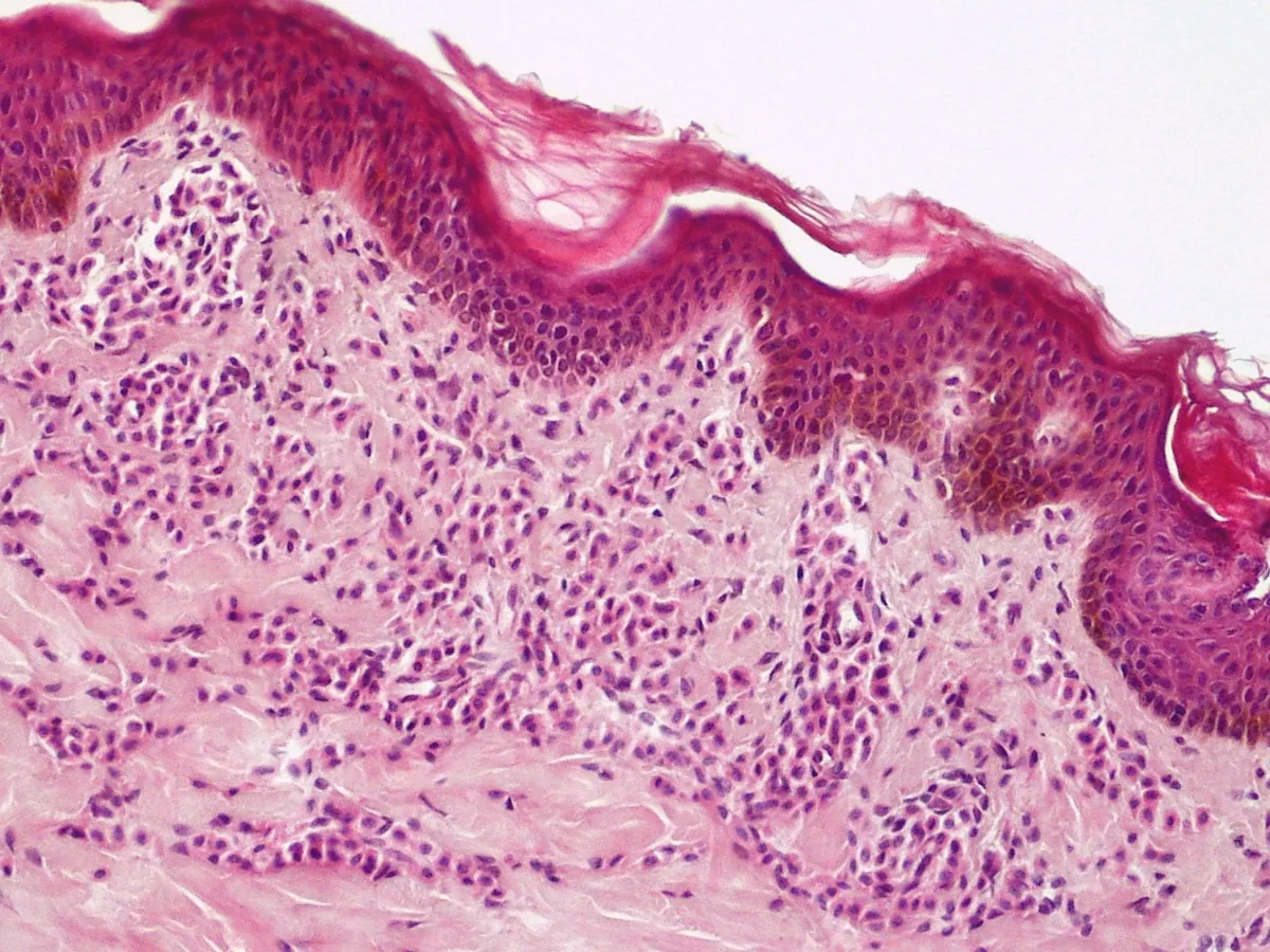
In the bone marrow (preferred) or other extracutaneous organs, multifocal dense aggregates of mast cells are found. Atypical or spindle-shaped mast cells and expression of the surface markers CD2 and/or CD25 on mast cells are typical.
Diagnosis
Systemic mastocytosis is confirmed when the major criterion and at least one minor criterion, or at least three of the four minor criteria, are met.
- Major criterion: multifocal dense aggregates of more than 15 mast cells in bone marrow or other extracutaneous organs (not the gastrointestinal tract, lymph nodes, liver or spleen).
- Minor criterion 1: more than 25% atypical or spindle-shaped mast cells in bone marrow sections or aspirate.
- Minor criterion 2: KIT mutation at codon 816 (usually D816V) in bone marrow, blood or other tissue.
- Minor criterion 3: expression of CD2, CD25 or both on mast cells from bone marrow or other extracutaneous organs.
- Minor criterion 4: baseline serum tryptase above 20 ng/mL; values above 11.4 ng/mL are considered elevated in most laboratories.
Baseline tryptase is elevated in systemic mastocytosis but usually normal in cutaneous mastocytosis and MCAS. A skin biopsy can show mast cell infiltrates but does not replace bone marrow biopsy for classification. In addition, mast cell mediators and their metabolites can be measured in urine or plasma (e.g. N-methylhistamine, prostaglandin D2, leukotriene E4). An increase in tryptase during an attack of at least 20% plus 2 ng/mL above baseline supports MCAS.
Keep learning in the app
Further reading (open access)
Cross-references
More topics: Allergology
Note: Learning content for medical education – not a treatment recommendation and no substitute for diagnosis or treatment decisions in individual cases. Treatment and management are deliberately not covered on this page.