Stevens-Johnson syndrome and toxic epidermal necrolysis
- Synonyms
- Lyell syndrome, TEN, SJS, epidermal necrolysis, SJS/TEN overlap
- Specialty
- Internal medicine · Allergology
- Images
- Clinical 2 · Histology 1
- Last updated
- 10/2026 · Dr. Pascal Bafteh
Contents
Images (3)
 Histology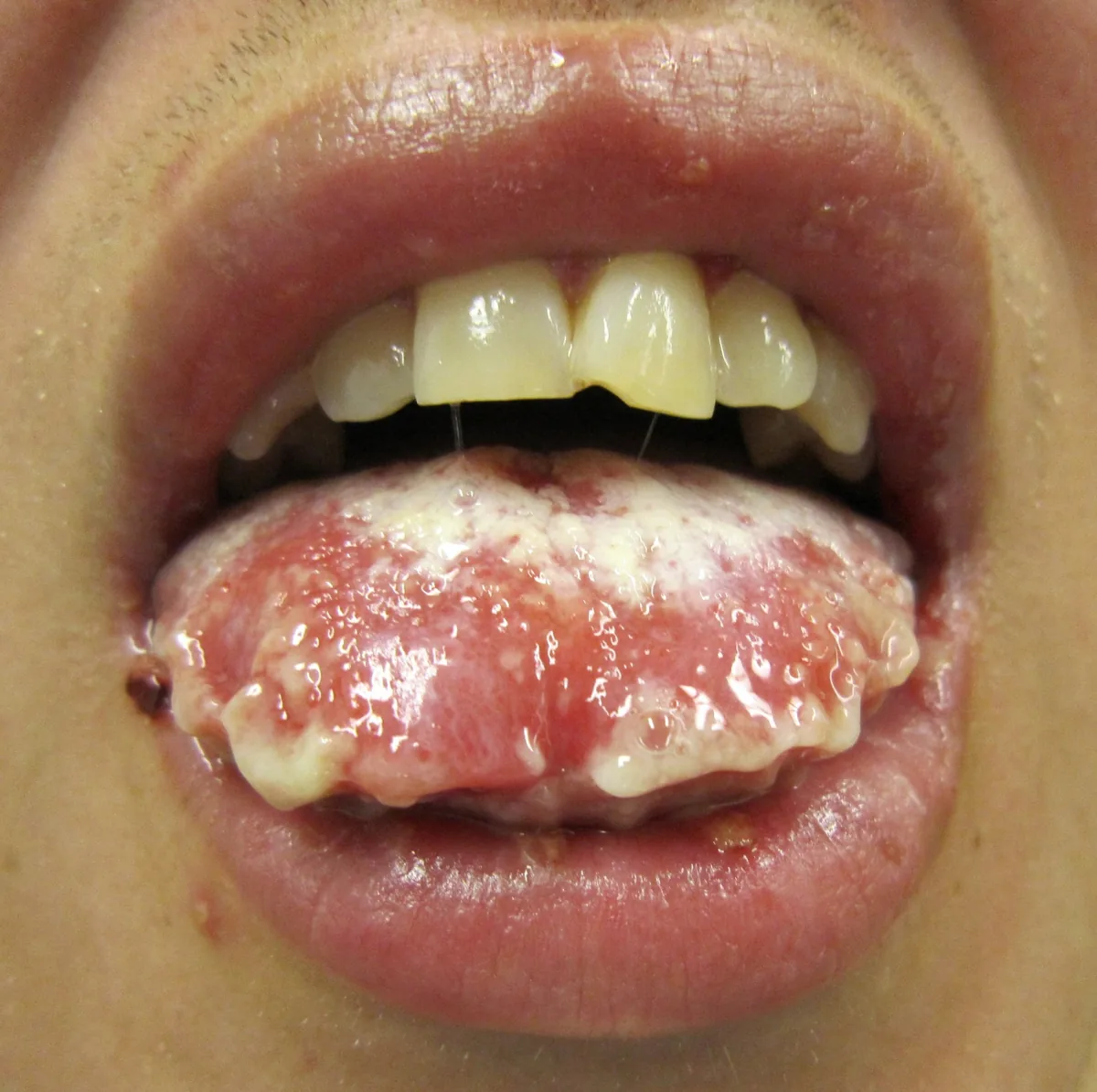
Definition
Stevens-Johnson syndrome (SJS) and toxic epidermal necrolysis (TEN, Lyell syndrome) are severe, acute hypersensitivity reactions of the skin and mucous membranes with extensive necrosis and detachment of the epidermis. Both belong to one disease spectrum and differ mainly in the extent of skin detachment.
Classification
- SJS: skin detachment of less than 10% of body surface area.
- SJS/TEN overlap: 10 to 30% of body surface area.
- TEN: more than 30% of body surface area.
Occurrence & epidemiology
About 2 to 9 people per million are affected. Incidence and severity are increased in bone marrow or stem cell recipients, in HIV infection with Pneumocystis jirovecii infection, in systemic lupus erythematosus and in other chronic systemic rheumatic diseases.
Aetiopathogenesis
- Drugs: the most common cause in adults; caused above all by sulfonamides, other anti-infectives (e.g. cephalosporins), antiepileptic drugs, NSAIDs and antiretroviral drugs.
- Other causes: infections, especially with Mycoplasma pneumoniae (the most likely cause in children) and graft-versus-host disease; rarely no cause is found.
- Pathogenesis: probably a cytotoxic T-cell reaction against drug antigens in keratinocytes, possibly favoured by altered clearance of reactive metabolites. CD8-positive T cells and natural killer cells release granulysin, whose concentration in blister fluid correlates with severity; interleukin-15 is increased. Fas- and Fas ligand-mediated keratinocyte death is also discussed.
- Genetic predisposition: strong associations with HLA alleles, e.g. HLA-B*58:01 with a uricostatic drug and HLA-A*31:01 and HLA-B*15:02 with carbamazepine.
Clinical features
- Onset: 1 to 3 weeks after starting the causative drug, with a prodrome of malaise, fever, headache, cough and keratoconjunctivitis.
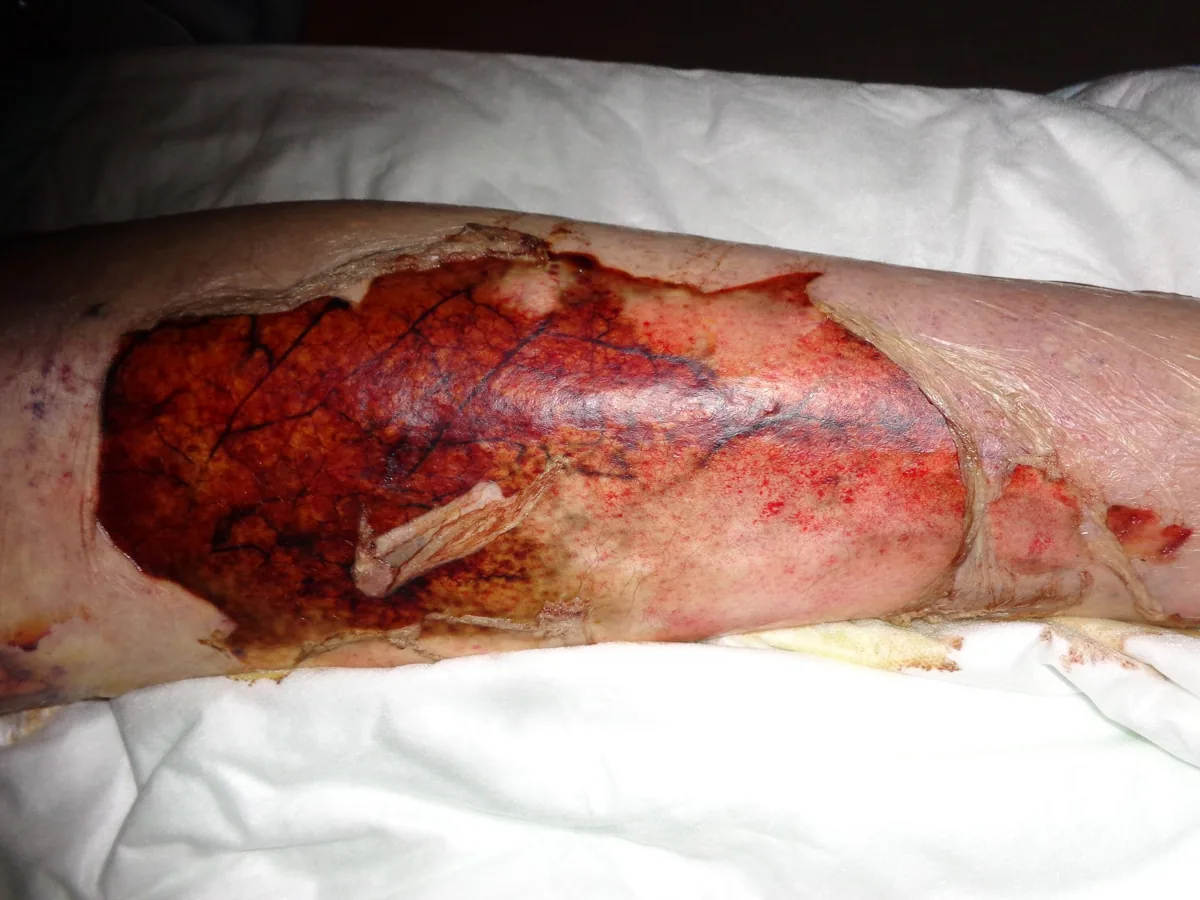
- Skin findings: sudden, often bull's-eye-like macules on the face, neck and upper trunk, rapidly also elsewhere; they coalesce into large flaccid bullae, and the epidermis sloughs within 1 to 3 days. In TEN, diffuse erythema may be the first sign.
- Nikolsky sign: positive; at pressure points large sheets of epidermis slide off, leaving weeping, painful, red areas. Palms, soles, nails and eyebrows may also be affected.
- Mucous membranes: painful oral erosions and crusts, keratoconjunctivitis and genital involvement (urethritis, phimosis, vaginal synechiae).
- Internal organs: sloughing of bronchial epithelium with cough, dyspnea, pneumonia, pulmonary edema and hypoxemia; also glomerulonephritis and hepatitis.
- Severe disease: similar to extensive burns, with massive fluid and electrolyte losses, a high risk of infection and multiorgan failure.
- Long-term sequelae: damage to skin, eyes and oral mucosa and psychological consequences.
Histology
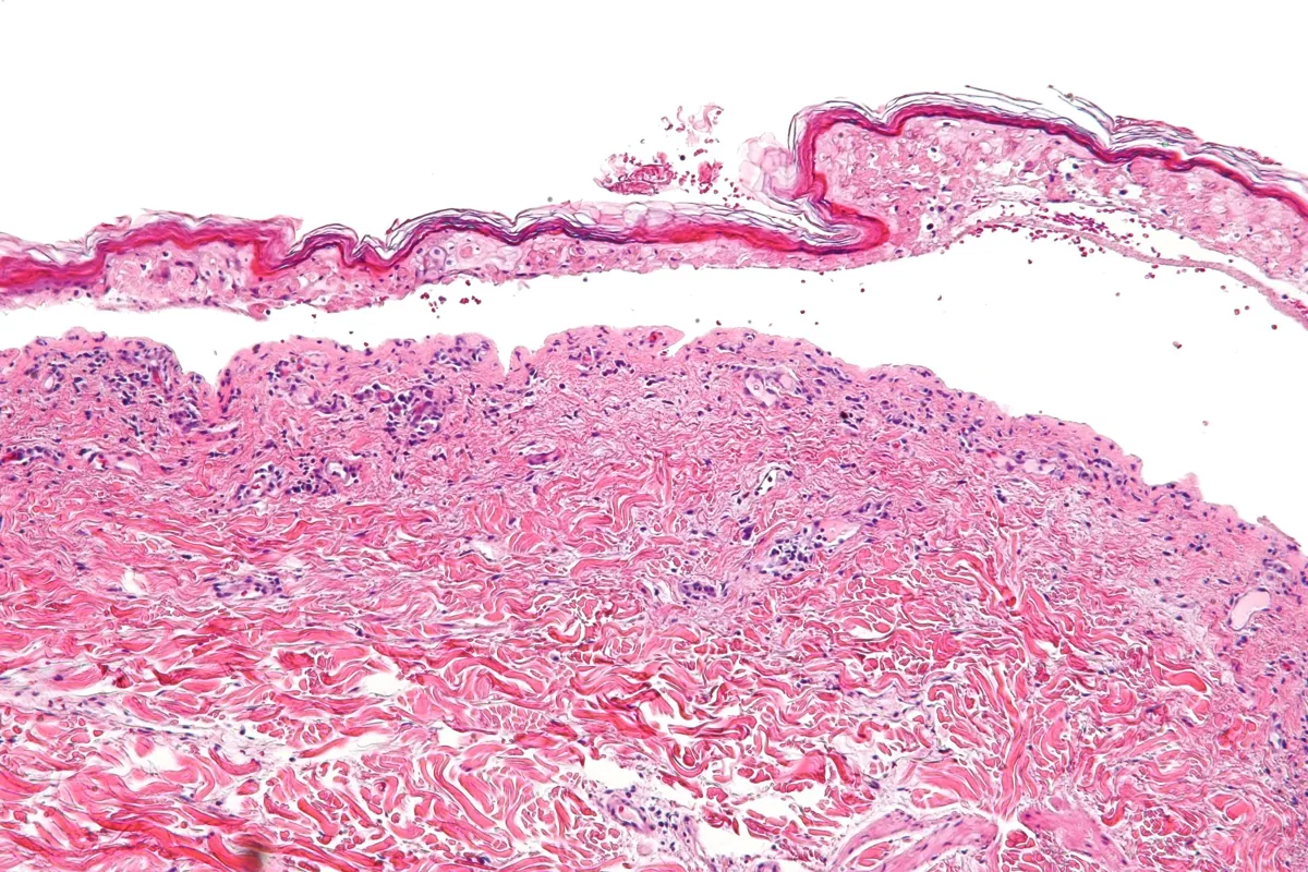
Histology of detached skin shows necrotic epithelium; this finding is an important distinguishing feature from other blistering disorders.
Diagnosis
- Clinical diagnosis: typical lesions, mucosal involvement, marked pain and rapid progression with a matching drug history.
- Skin biopsy: frequently used for confirmation; demonstrates epidermal necrosis.
- Extent of detachment: determination of the affected body surface area to classify as SJS, overlap or TEN.
Keep learning in the app
Further reading (open access)
Cross-references
Note: Learning content for medical education – not a treatment recommendation and no substitute for diagnosis or treatment decisions in individual cases. Treatment and management are deliberately not covered on this page.