Hypercalcemia
Board exam relevance: in 10 of 105 exam reports · rank 27- Synonyms
- high calcium, high blood calcium, hypercalcaemia of malignancy, hyperparathyroidism
- Specialty
- Internal medicine · Electrolytes & acid–base
- Images
- Gross specimen 1 · Histology 1
- Last updated
- 10/2026 · Dr. Pascal Bafteh
Contents
Images (2)
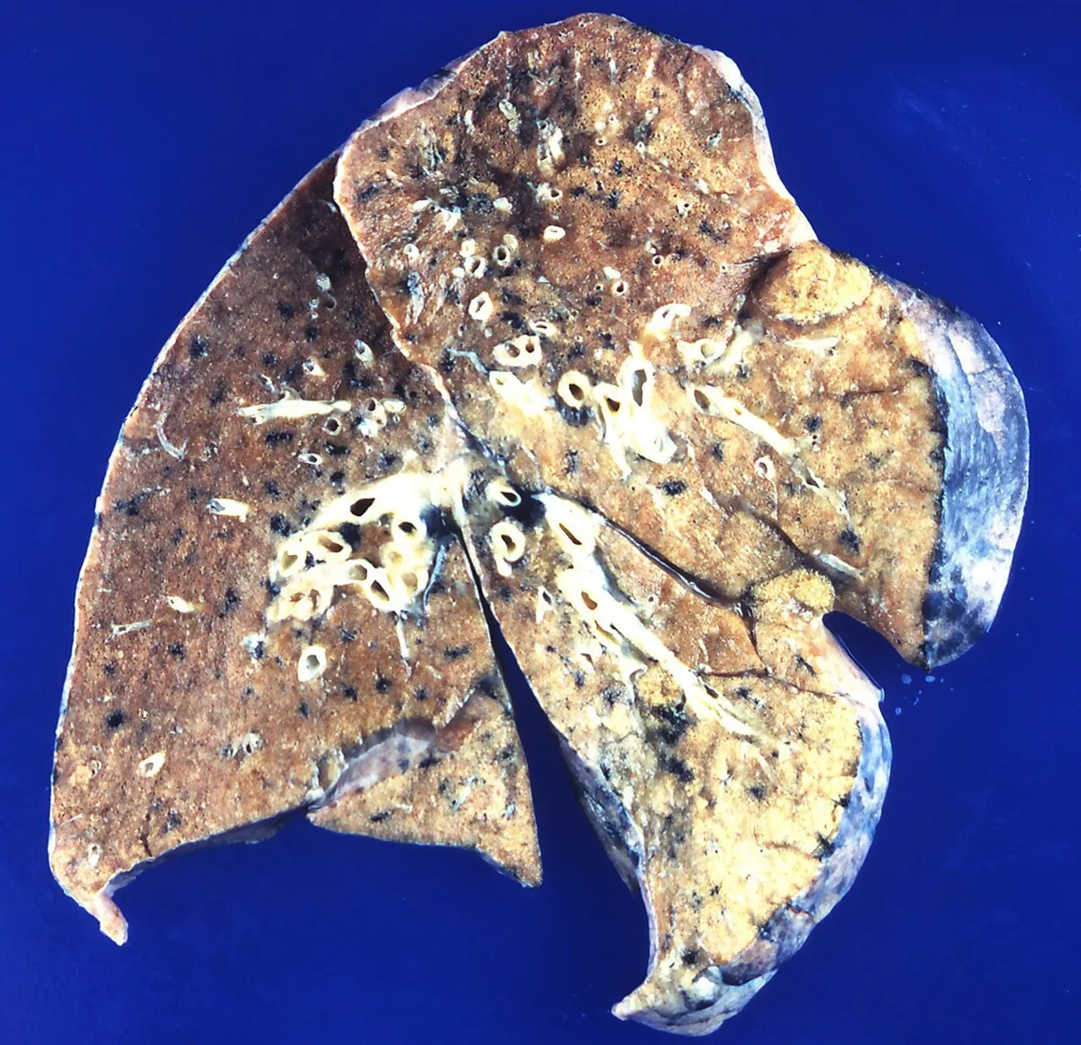 Gross specimen
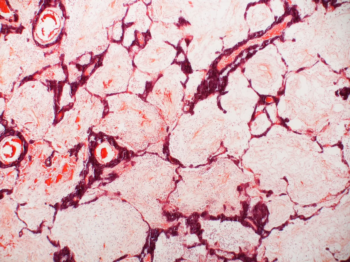 Histology
Definition
Hypercalcemia is defined as a total serum calcium above 2.60 mmol/L or an ionised calcium above 1.30 mmol/L. It results mainly from increased bone resorption, less often from increased intestinal absorption or reduced renal excretion. The two most common causes are primary hyperparathyroidism and cancer. Hypercalcemia is often discovered incidentally on routine laboratory testing.
Classification
- By severity (total calcium): mild about 2.60 to 2.97 mmol/L, moderate about 2.99 to 3.47 mmol/L, severe above 3.49 mmol/L.
- By mechanism: PTH-dependent forms (primary, secondary and tertiary hyperparathyroidism, familial hypocalciuric hypercalcemia) with high or high-normal parathyroid hormone, and PTH-independent forms (cancer, vitamin D excess, granulomatous diseases and others) with suppressed parathyroid hormone.
Occurrence & epidemiology
Primary hyperparathyroidism is probably the most common cause, especially in outpatients. Its incidence rises with age and is higher in women, especially after menopause. Cancer-related hypercalcemia is found mainly in hospital patients.
Aetiopathogenesis
Parathyroid hormone (PTH) mobilises calcium and phosphate from bone, increases distal calcium reabsorption, reduces proximal phosphate reabsorption and promotes the formation of calcitriol, which increases intestinal calcium absorption.
- Primary hyperparathyroidism: in about 85% a parathyroid adenoma, in about 15% hyperplasia of two or more glands, in less than 1% a carcinoma. Familial forms occur as part of multiple endocrine neoplasia.
- Secondary and tertiary hyperparathyroidism: mainly in advanced chronic kidney disease; in tertiary hyperparathyroidism PTH is secreted autonomously after long-standing stimulation.
- Humoral hypercalcemia of malignancy: production of PTH-related peptide (PTHrP), most often with squamous cell carcinomas and renal cell, breast, prostate and ovarian carcinoma.
- Osteolytic hypercalcemia of malignancy: bone metastases from solid tumors (e.g. breast, prostate, non-small cell lung cancer) and hematological malignancies, above all multiple myeloma.
- Vitamin D-mediated: vitamin D toxicity, calcitriol production in granulomas (sarcoidosis, tuberculosis, histoplasmosis and others) or in lymphomas.
- Other causes: immobilisation (especially in Paget disease or multiple fractures), hyperthyroidism, adrenal insufficiency, vitamin A toxicity, milk-alkali syndrome due to calcium carbonate antacids, lithium or theophylline toxicity and distally acting saluretics.
- Familial hypocalciuric hypercalcemia (FHH): autosomal dominant, usually an inactivating mutation in the calcium-sensing receptor gene (CASR, less often GNA11 or AP2S1); usually asymptomatic, with hypocalciuria and often hypermagnesemia.
Clinical features
- Gastrointestinal tract (mild forms often asymptomatic): constipation, loss of appetite, nausea, vomiting, abdominal pain and ileus.
- Kidney: polyuria, nocturia and polydipsia due to impaired concentrating ability; nephrolithiasis with hypercalciuria; less often acute kidney injury or nephrocalcinosis.
- Nervous system and mind: from about 3.00 mmol/L emotional lability, confusion, delirium, psychosis, stupor and coma; also muscle weakness.
- Heart: in severe hypercalcemia a shortened QTc interval and arrhythmias, especially with digitalis.
- Extreme hypercalcemia: above about 4.50 mmol/L, shock and renal failure may occur.
Diagnosis
Laboratory tests
- Total and ionised calcium: high serum protein raises total calcium artefactually, low protein masks true hypercalcemia; ionised calcium is then decisive.
- Parathyroid hormone (intact): high or high-normal in PTH-dependent forms; in PTH-independent causes usually below 2.1 pmol/L. A raised intact PTH with an inappropriately high or high-normal calcium confirms hyperparathyroidism.
- Phosphate and alkaline phosphatase: low phosphate suggests hyperparathyroidism; alkaline phosphatase is often raised when bone turnover is increased.
- Calcium level: without an evident cause, values below 2.75 mmol/L suggest hyperparathyroidism or another benign cause, values above 3.25 mmol/L suggest cancer. In hyperparathyroidism total calcium rarely exceeds 3 mmol/L.
- Further baseline tests: electrolytes, urea, creatinine, 25-hydroxyvitamin D and serum immunoelectrophoresis. Together with history, examination, chest X-ray, calcium, phosphate, PTH and alkaline phosphatase, these identify the cause in at least 95% of cases. Targeted additions are 1,25-dihydroxyvitamin D (raised e.g. in sarcoidosis and some lymphomas) and PTHrP (humoral hypercalcemia of malignancy).
- 24-hour urinary calcium: low in FHH, normal or high in hyperparathyroidism and cancer. Fractional calcium excretion is below 1% in FHH and 1 to 4% in primary hyperparathyroidism.
- Pointers to myeloma: anemia, azotaemia and hypercalcemia or monoclonal gammopathy; confirmed in bone marrow.
Imaging
- Chest X-ray: reveals most granulomatous diseases, lung cancer and lytic lesions and Paget changes in the scapulae, ribs and thoracic spine.
- Skeletal radiographs: in symptomatic hyperparathyroidism (osteitis fibrosa cystica), bone cysts, a heterogeneous skull appearance and subperiosteal resorption of the finger phalanges and distal clavicles.
- Localisation of the parathyroid glands: high-resolution ultrasound, CT, MRI and technetium-99m sestamibi scintigraphy, which is particularly sensitive for solitary adenomas.
Keep learning in the app
Further reading (open access)
Cross-references
Note: Learning content for medical education – not a treatment recommendation and no substitute for diagnosis or treatment decisions in individual cases. Treatment and management are deliberately not covered on this page.