Hypocalcemia
Board exam relevance: in 1 of 105 exam reports · rank 181- Synonyms
- low calcium, low blood calcium, tetany, hypoparathyroidism
- Specialty
- Internal medicine · Electrolytes & acid–base
- Images
- Clinical 1 · ECG 1
- Last updated
- 10/2026 · Dr. Pascal Bafteh
Contents
Images (2)
 ECG
ECGDefinition
Hypocalcemia is defined as a total serum calcium below 2.20 mmol/L with normal plasma proteins or an ionised calcium below 1.17 mmol/L. Because low protein levels lower total but not ionised calcium, calcium is always interpreted together with albumin. Hypocalcemia due solely to hypoproteinemia causes no symptoms and is termed factitious hypocalcemia.
Aetiopathogenesis
- Hypoparathyroidism: postoperative damage to the parathyroid glands in the neck (symptoms usually 24 to 48 hours postoperatively, occasionally only after months or years), autoimmune, as part of polyglandular autoimmune syndromes or congenital (e.g. DiGeorge syndrome). Hypocalcemia and hyperphosphatemia are typical.
- Pseudohypoparathyroidism: lack of PTH action on kidney and bone despite high PTH levels. Type Ia (Albright hereditary osteodystrophy, GNAS mutation) is associated with short stature, a round face, shortened metacarpals and metatarsals and basal ganglia calcification.
- Vitamin D deficiency: low sun exposure, inadequate intake, malabsorption or liver disease, and certain antiepileptic drugs that alter vitamin D metabolism; less often inherited vitamin D-dependent rickets.
- Kidney disease: reduced calcitriol formation in renal failure with hyperphosphatemia, and renal tubular acidoses.
- Magnesium depletion: especially below 0.5 mmol/L it causes relative PTH insufficiency and reduced tissue responsiveness to PTH.
- Other causes: acute pancreatitis (calcium binding by fatty acids), hungry bone syndrome after elimination of marked hyperparathyroidism, septic shock, more than 10 units of citrated blood and EDTA-containing contrast agents. Marked alkalosis lowers ionised calcium.
Clinical features
- Often asymptomatic: especially in mild or chronic hypocalcemia.
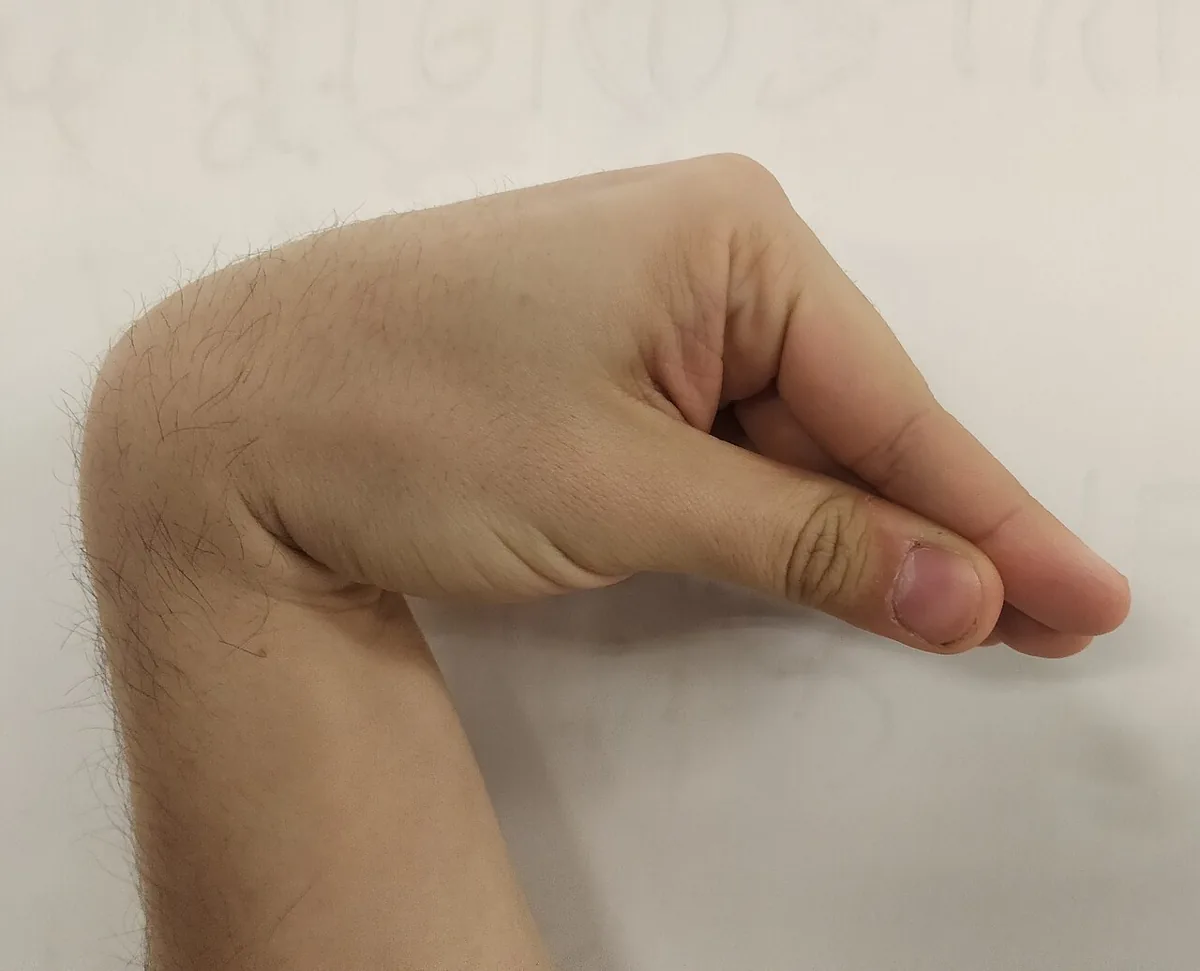
- Neuromuscular irritability: paraesthesia of the lips, tongue, fingers and feet, muscle cramps of the back and legs, painful carpopedal spasm and spasm of the facial muscles (tetany).
- Severe hypocalcemia (below about 1.75 mmol/L): hyperreflexia, laryngospasm and generalised seizures.
- Latent tetany (about 1.75 to 2.20 mmol/L): demonstrated by Chvostek's sign (twitching of the facial muscles when the facial nerve is tapped in front of the ear; positive in up to 10% of healthy people) and Trousseau's sign (carpal spasm after 3 minutes of a blood pressure cuff inflated to 20 mmHg above systolic pressure).
- Chronic hypocalcemia: diffuse encephalopathy, depression or psychosis, occasionally papilledema, dry scaly skin, brittle nails, coarse hair, cataract and candida infections.
Diagnosis
- Calcium and albumin: total calcium with estimation of the ionised fraction from albumin; if suspected, direct measurement of ionised calcium.
- Basic laboratory tests: creatinine and urea, phosphate, magnesium and alkaline phosphatase.
- Intact parathyroid hormone: a low or low-normal PTH is inappropriate in hypocalcemia and suggests hypoparathyroidism (then phosphate high, alkaline phosphatase normal); an undetectable PTH suggests idiopathic hypoparathyroidism. A high PTH suggests pseudohypoparathyroidism or a disorder of vitamin D metabolism.
- Vitamin D: 25-hydroxy- and 1,25-dihydroxyvitamin D; in vitamin D deficiency typically low phosphate and high alkaline phosphatase.
- Urinary cAMP and urinary phosphate: if pseudohypoparathyroidism is suspected.
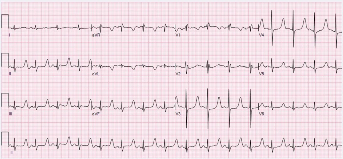
- ECG: in severe hypocalcemia prolongation of the QTc interval and ST segment, T-wave changes, occasionally arrhythmias or heart block.
- X-ray: signs of rickets or osteomalacia in vitamin D deficiency.
Keep learning in the app
Further reading (open access)
Cross-references
Note: Learning content for medical education – not a treatment recommendation and no substitute for diagnosis or treatment decisions in individual cases. Treatment and management are deliberately not covered on this page.