Chondrodermatitis nodularis helicis
Exam relevance: in 1 of more than 50 board exam reports · rank 93
- Synonyms
- Morbus Winkler, Chondrodermatitis nodularis chronica helicis, schmerzhaftes Ohrknötchen, chondrodermatitis nodularis, Winkler disease, painful ear nodule
- Specialty
- Dermatology · Regional & special dermatoses
- Images
- Clinical 2
- Last updated
- 10/2026 · Dr. Pascal Bafteh
Contents
Images (2)
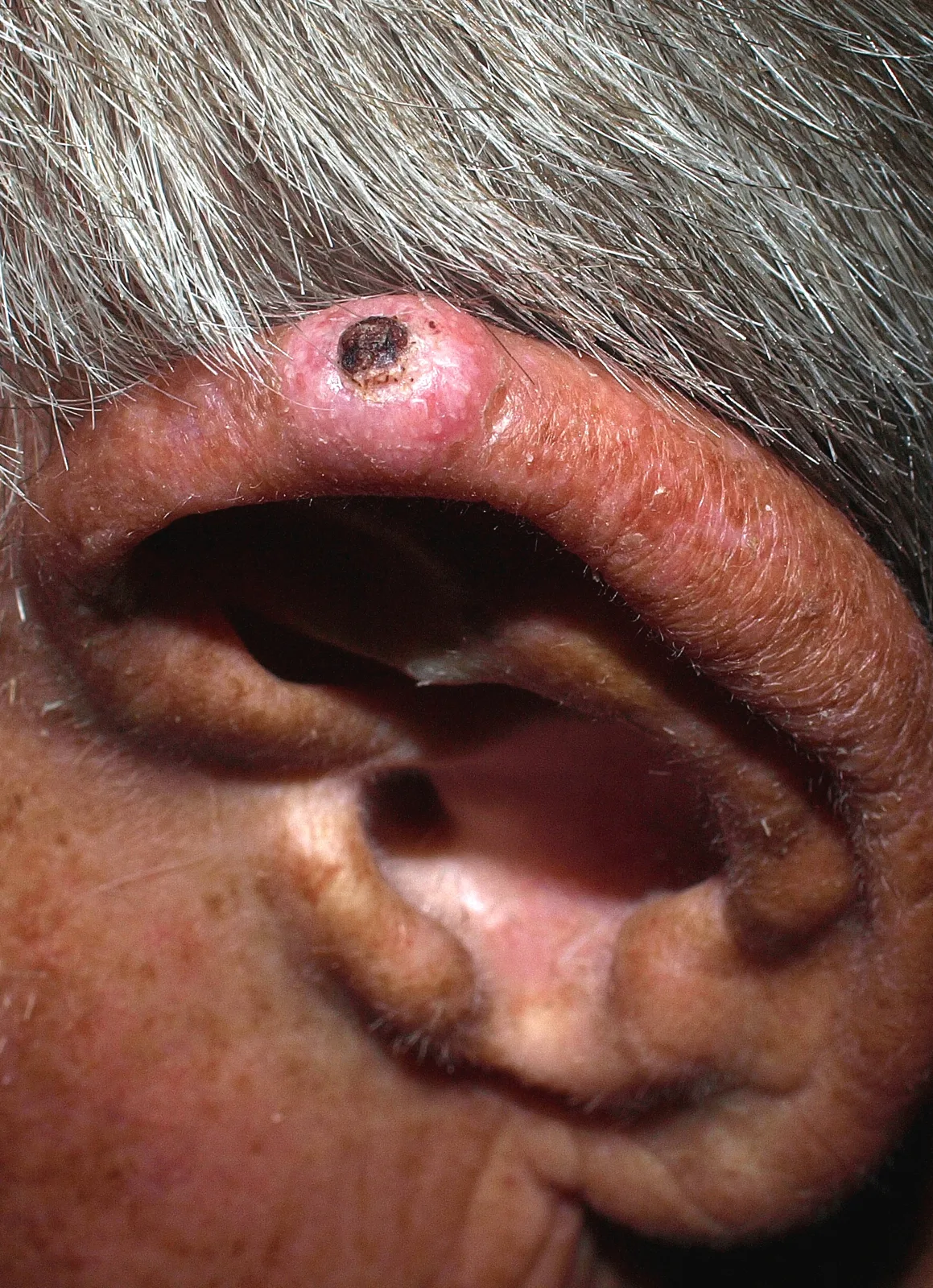
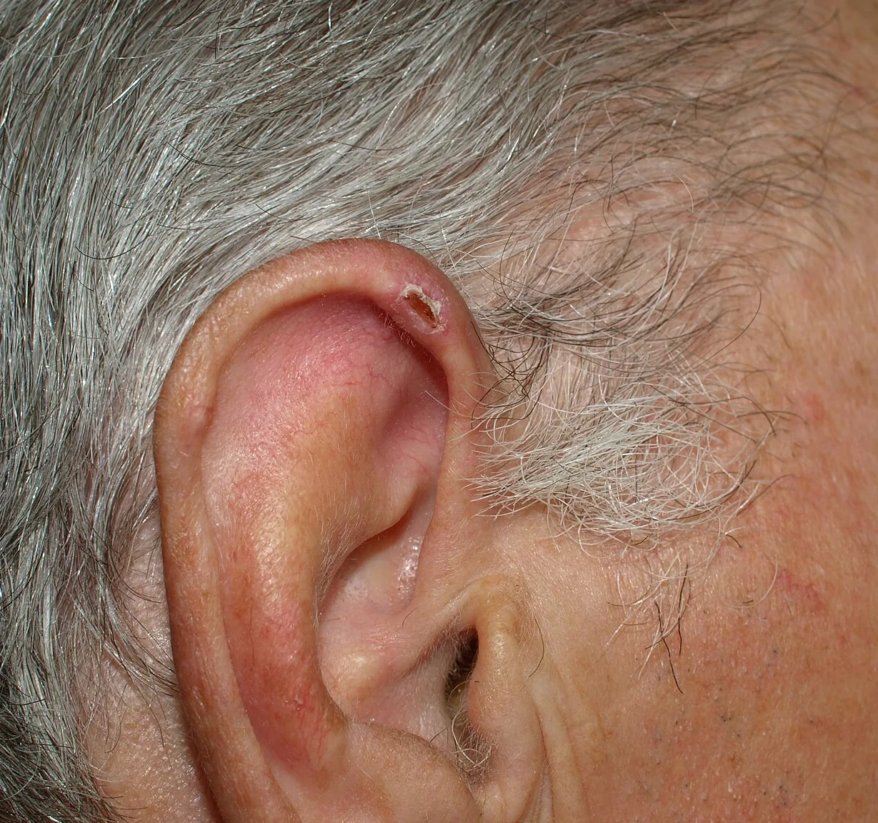
Definition
Chondrodermatitis nodularis helicis (Winkler disease, after its first description in 1915) is a common inflammatory condition of the skin and cartilage of the helix or antihelix of the ear.
Occurrence & epidemiology
It occurs mainly in fair-skinned middle-aged and older men; only 10–35% of reported cases are in women, and it is rare in children. The exact cause is unknown; contributing factors are repeated pressure with reduced blood supply (mostly on the side slept on), cold and sun, pressure from headphones, mobile devices or hearing aids, and connective tissue diseases such as lupus erythematosus, dermatomyositis and scleroderma.
Clinical features
- A solitary, firm, oval nodule of 4–6 mm with a central crust and surrounding erythema.
- In men usually on the helix, in women more often on the antihelix; typically unilateral on the sleeping side, rarely bilateral.
- Painful and tender; night pain often prevents sleeping on that side. The nodule may bleed or discharge a little scaly material.
Diagnosis
- Usually a clinical diagnosis based on the typical site on the helix or antihelix and the history of pain and tenderness.
- Occasionally histological confirmation is needed.
Treatment principles
- The basic principle is relief of pressure on the affected ear, for example with a special pillow with a cut-out, which alone can lead to improvement.
- Intralesional glucocorticoids are used as an adjunct.
- If the lesion persists, surgical removal of the affected cartilage is the most effective option; histology also serves to exclude squamous or basal cell carcinoma, and recurrences are possible.
Specific treatment regimens with drugs and doses are covered in the DermaFuchs app.
Keep learning in the app
References (selection)
Cross-references
Keep learning
Keep learning with flashcards, exam questions and spot diagnoses in the DermaFuchs app – free, in your browser or as an app.
Related pages: Condylomata acuminata (genital warts) · Paraneoplastic dermatoses · Dermatoses of pregnancy
Note: This page is intended for medical education and does not replace diagnosis or treatment decisions in individual cases.