Uterine inversion
- Specialty
- Obstetrics · Birth & puerperium
- Images
- Clinical 1
- Last updated
- 10/2026 · Dr. Pascal Bafteh
Contents
Images (1)
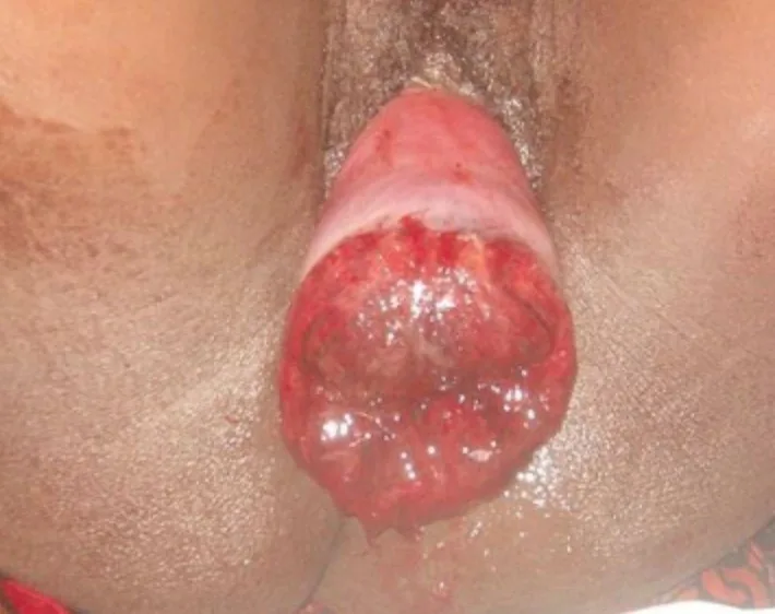
Definition
- In uterine inversion the fundus descends into the uterine cavity, through the cervix or beyond the introitus; about 85 per cent of cases occur postpartum, typically in the third stage of labour.
Classification
- Puerperal (obstetric) inversion, which may be acute or chronic, is distinguished from non-puerperal inversion, which is mostly chronic and related to uterine tumours, most commonly leiomyomas (56%) and less often malignancies (32%).
Occurrence & epidemiology
Epidemiology
- Puerperal uterine inversion is a rare, life-threatening obstetric emergency; a higher incidence has been reported after caesarean section.
Aetiopathogenesis
Aetiology and risk factors
- The commonest cause is excessive cord traction during birth of the placenta; contributing factors are excessive fundal pressure in the third stage, uterine atony and placenta accreta.
Clinical features
Clinical features and complications
- Puerperal inversion is closely associated with postpartum haemorrhage; blood loss often requires transfusion and may progress to hypovolaemic shock and maternal death.
- On abdominal examination the fundus is not palpable or lower than expected, and the inverted fundus may be visible at or beyond the introitus.
Diagnosis
- The diagnosis of puerperal inversion is clinical.
- Ultrasound shows a U-shaped configuration in the sagittal plane and a target sign in the transverse plane; in the non-puerperal form, ultrasound and MRI enable the diagnosis, and distinguishing it from tumours or prolapse can be difficult.
Keep learning in the app
Further reading (selection)
Cross-references
Note: Learning content from the GynFuchs app (flashcards, exam questions, image cases) for medical education – not a treatment recommendation and no substitute for diagnosis or treatment decisions in individual cases. Treatment and management are deliberately not covered on this page.