Cervical dysplasia (CIN)

Exam relevance: in 44 of 197 board exam reports · rank 8

Synonyms
cervical dysplasia, CIN, abnormal smear, cervical precancer
Specialty
Gynaecology · Cervix & HPV
Images
Colposcopy 10 · Clinical 2
Exam relevance
44 of 197 reports · rank 8
In the app
4 flashcards · GynFuchs
Last updated
10/2026 · Dr. Pascal Bafteh
Contents
  1. Images (12)
  2. Definition
  3. Classification
  4. Aetiopathogenesis
  5. Clinical features
  6. Diagnosis
  7. Keep learning in the app
  8. Further reading (selection)
  9. Cross-references

Images (12)

Cervical dysplasia (CIN) – Colposcopy: Type 3 transformation zone — the squamocolumnar junction is not visibleColposcopy
Type 3 transformation zone — the squamocolumnar junction is not visibleImage: Vinokurov MA, Minaeva AV, Leshkina GV et al., Front Reprod Health 2025 · CC BY 4.0 · Source · cropped
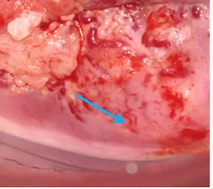
Cervical dysplasia (CIN) – Clinical: Atypical, bizarre vessels over an exophytic, necrotic cervical lesion
Atypical, bizarre vessels over an exophytic, necrotic cervical lesionImage: Zhang Y, Li H, Li X et al., Front Oncol 2023 · CC BY 4.0 · Source · cropped
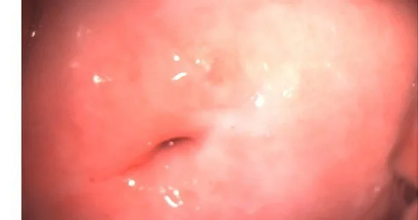
Cervical dysplasia (CIN) – Colposcopy: Ectopy/ectropion in a nullipara — red columnar epithelium around the os with a sharp squamous borderColposcopy
Ectopy/ectropion in a nullipara — red columnar epithelium around the os with a sharp squamous borderImage: GynaeImages (Wikimedia Commons) · CC BY-SA 4.0 · Source
Cervical dysplasia (CIN) – Clinical
Image: Vinokurov MA, Minaeva AV, Leshkina GV et al., Front Reprod Health 2025 · CC BY 4.0 · Source
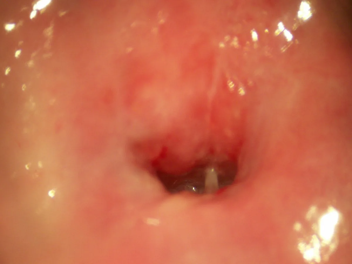
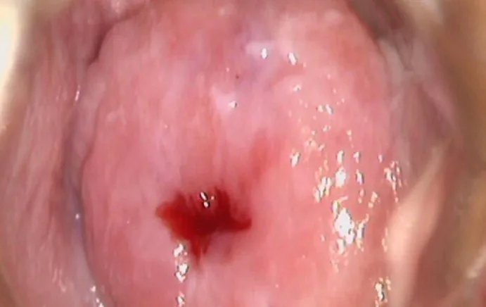
Cervical dysplasia (CIN) – Colposcopy: Close-up of the external cervical os with cervical mucusColposcopy
Close-up of the external cervical os with cervical mucusImage: Bobjgalindo (Wikimedia Commons) · CC BY-SA 4.0 · Source
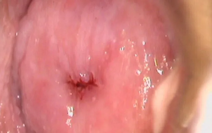
Cervical dysplasia (CIN) – Colposcopy: Cervix before acetic acid — the baseline colposcopic view (Photo: Milic N, Varnicic Lojanica M, Ivanovic S et al., Diagnostics (Basel) 2026, open access, CC BY 4.0)Colposcopy
Cervix before acetic acid — the baseline colposcopic view (Photo: Milic N, Varnicic Lojanica M, Ivanovic S et al., Diagnostics (Basel) 2026, open access, CC BY 4.0)Image: Milic N, Varnicic Lojanica M, Ivanovic S et al., Diagnostics (Basel) 2026 · CC BY 4.0 · Source · cropped
Cervical dysplasia (CIN) – Colposcopy: The same cervix one minute after 3% acetic acid — only faint acetowhitening around the os (Photo: Milic N, Varnicic Lojanica M, Ivanovic S et al., Diagnostics (Basel) 2026, open access, …Colposcopy
The same cervix one minute after 3% acetic acid — only faint acetowhitening around the os (Photo: Milic N, Varnicic Lojanica M, Ivanovic S et al., Diagnostics (Basel) 2026, open access, …Image: Milic N, Varnicic Lojanica M, Ivanovic S et al., Diagnostics (Basel) 2026 · CC BY 4.0 · Source · cropped
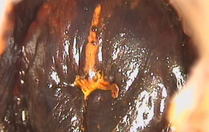
Cervical dysplasia (CIN) – Colposcopy: Lugol test: a mahogany-brown iodine-positive cervix with a clearly yellow iodine-negative area at the os (Photo: Milic N, Varnicic Lojanica M, Ivanovic S et al., Diagnostics (Basel) …Colposcopy
Lugol test: a mahogany-brown iodine-positive cervix with a clearly yellow iodine-negative area at the os (Photo: Milic N, Varnicic Lojanica M, Ivanovic S et al., Diagnostics (Basel) …Image: Milic N, Varnicic Lojanica M, Ivanovic S et al., Diagnostics (Basel) 2026 · CC BY 4.0 · Source · cropped
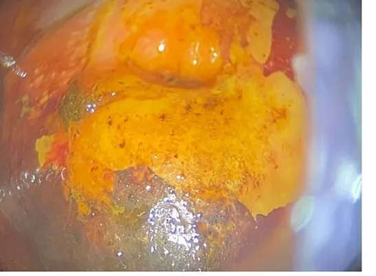
Cervical dysplasia (CIN) – Colposcopy: Iodine-negative (yellow) cervix after Lugol solution — a Schiller-positive finding (Photo: NCI DES-Atlas (US-Bundesbehörde, PD), Wikimedia Commons, Public domain)Colposcopy
Iodine-negative (yellow) cervix after Lugol solution — a Schiller-positive finding (Photo: NCI DES-Atlas (US-Bundesbehörde, PD), Wikimedia Commons, Public domain)Image: NCI DES-Atlas (US-Bundesbehörde, PD) (Wikimedia Commons) · Public domain · Source
Cervical dysplasia (CIN) – Colposcopy: Dense, sharply demarcated acetowhite epithelium — a grade 2 major changeColposcopy
Dense, sharply demarcated acetowhite epithelium — a grade 2 major changeImage: Vinokurov MA, Minaeva AV, Leshkina GV et al., Front Reprod Health 2025 · CC BY 4.0 · Source · cropped
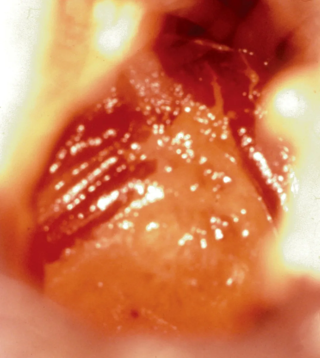
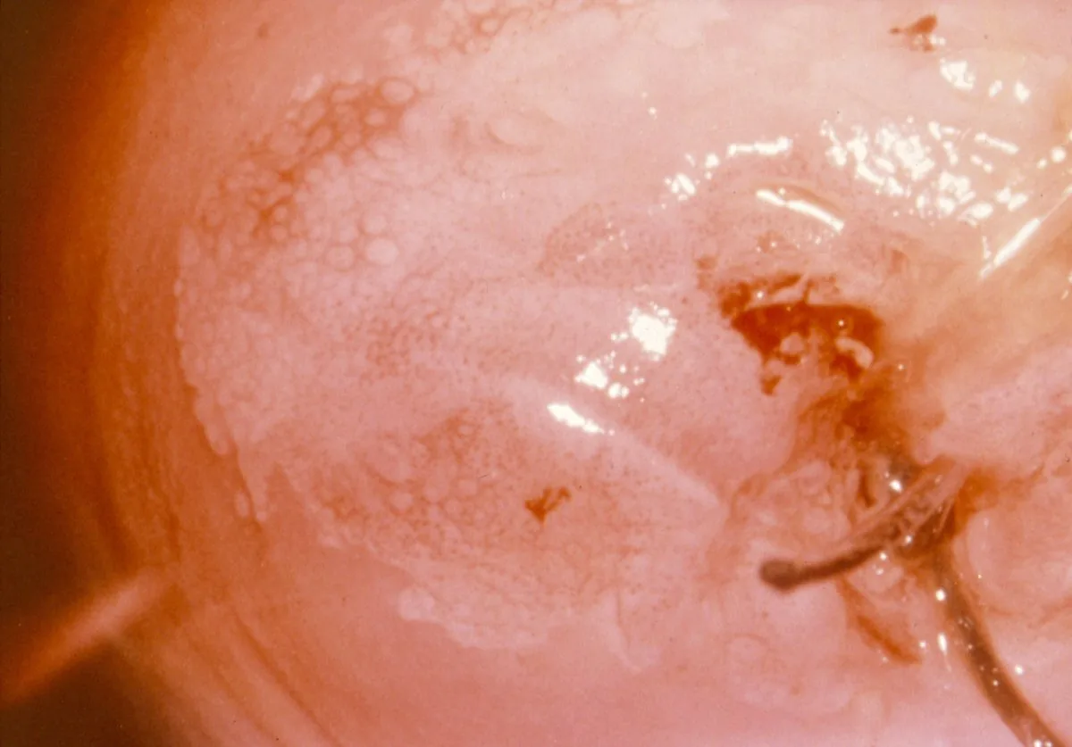
Cervical dysplasia (CIN) – Colposcopy: Coarse mosaic on the cervix — a major colposcopic change (Photo: NCI, 'An Atlas of Findings of Human Females After Intrauterine Exposure to DES' (US-Bundesbehörde, PD), Wikimedia …Colposcopy
Coarse mosaic on the cervix — a major colposcopic change (Photo: NCI, 'An Atlas of Findings of Human Females After Intrauterine Exposure to DES' (US-Bundesbehörde, PD), Wikimedia …Image: NCI, 'An Atlas of Findings of Human Females After Intrauterine Exposure to DES' (US-Bundesbehörde, PD) (Wikimedia Commons) · Public domain · Source
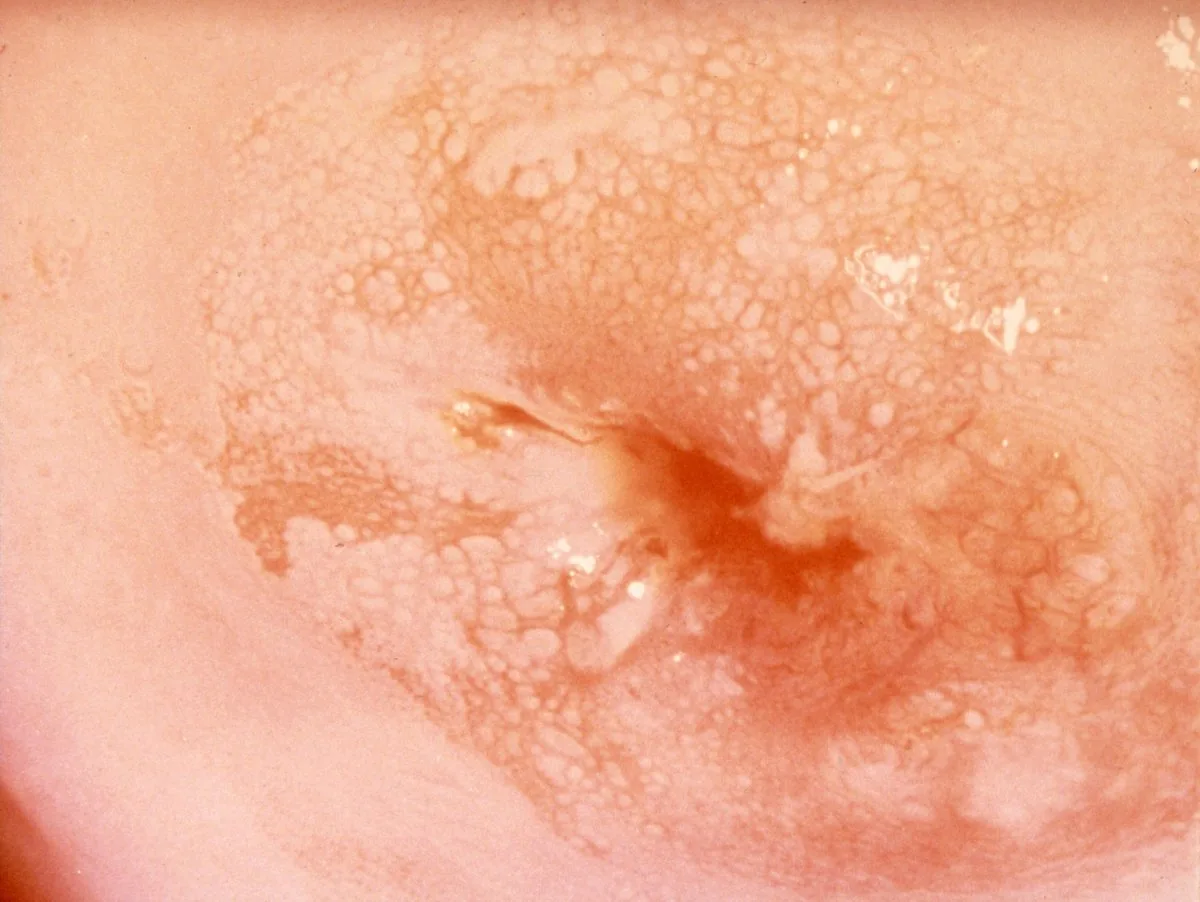
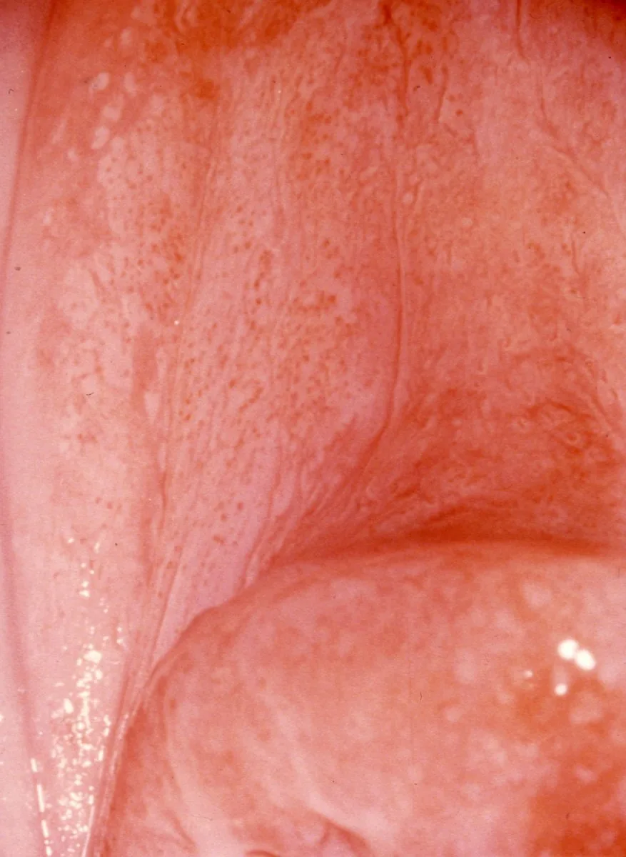
Cervical dysplasia (CIN) – Colposcopy: Fine, regular punctation — a minor change (Photo: NCI DES-Atlas (US-Bundesbehörde, PD), Wikimedia Commons, Public domain)Colposcopy
Fine, regular punctation — a minor change (Photo: NCI DES-Atlas (US-Bundesbehörde, PD), Wikimedia Commons, Public domain)Image: NCI DES-Atlas (US-Bundesbehörde, PD) (Wikimedia Commons) · Public domain · Source
1 / 12

Definition

What the transformation zone is

  • The TZ is the area between the original and the current squamocolumnar junction — through squamous metaplasia virtually every dysplasia arises there.

Classification

Three types

  • Type 1: TZ entirely ectocervical and fully visible.
  • Type 2: TZ extends endocervically but is fully visible with instruments.
  • Type 3: TZ endocervical and not fully visible — colposcopy inadequate.

Munich Nomenclature III (cytology)

  • Pap IIID1: cytologically mild dysplasia, histological correlate CIN I.
  • Pap IIID2: cytologically moderate dysplasia, histological correlate CIN II.
  • Pap IVa: severe dysplasia or carcinoma in situ, correlate CIN III.
  • Pap IVb: as IVa, plus invasion cannot be excluded.
  • Pap V: cells of an invasive carcinoma.

Aetiopathogenesis

Its migration through life

  • Before menarche: the junction lies endocervically, i.e. inside.
  • Young woman of reproductive age: oestrogen and eversion place it ectopically on the portio, columnar epithelium visible.

Clinical features

More facts from the study questions

  • Postmenopausally, the squamocolumnar junction recedes into the endocervical canal.
  • This often leads to a type 3 transformation zone.
  • A type 3 transformation zone extends endocervically and is by definition not completely visible.
  • A colposcopy is considered inadequate in this case.
  • In a type 1 transformation zone, the entire zone is located on the ectocervix.
  • It is therefore completely visible on examination.
  • In young, reproductive-age women, oestrogen influence leads to an ectropion.
  • This displaces columnar epithelium onto the portio surface, making the junction ectopic.
  • As the endocervical component of a TZ type 3 is not visible, tissue sampling from the cervical canal is required for an abnormal finding.
  • The finding Pap group IIID1 describes mild dysplasia cytologically.

Diagnosis

Acetic acid test

  • Concentration: 3–5 % acetic acid.
  • Mechanism: reversible coagulation of nuclear proteins — nucleus-rich dysplastic areas turn white.
  • Hence dysplastic, nucleus-rich epithelium turns acetowhite.
  • Prognostically important: the speed and density of the whitening — fast and dense suggests high grade.

Schiller iodine test (Lugol)

  • What stains is mature, glycogen-rich non-keratinised squamous epithelium — it turns mahogany brown.
  • Therefore iodine-negative (pale) are: dysplastic epithelium, columnar epithelium and atrophic epithelium.

Minor changes (grade 1)

  • Faintly acetowhite area with an ill-defined, geographic border.
  • Fine punctation and fine mosaic — small, regular vessel pattern.
  • Slow and weak acetic reaction that fades quickly.
  • Histological correlate usually CIN I.

Major changes (grade 2)

  • Dense, rapidly appearing acetowhitening with a sharp border.
  • Prominent, cuffed gland openings with a white rim.
  • Histological correlate usually CIN II or CIN III.

Suspicion of invasion

  • Atypical vessels with calibre changes and abrupt vessel endings are the decisive signs.
  • Plus: exophytic or ulcerated lesion, friable tissue that bleeds easily, irregular surface.
  • Consequence: targeted biopsy and start staging, do not merely observe.

Typical imaging findings

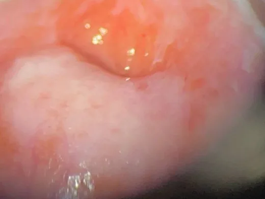
  • Colposcopy: A dense, sharply demarcated, strongly white area lies at the transformation zone and extends to the os. The whitening appears quickly and persists; the margins are smoothly defined, not frayed.
  • Colposcopy: A closed, dense white rim lights up around the os and is sharply demarcated from the surrounding pink epithelium. The whitening is strong and persistent, and no vascular pattern can be seen within it.
  • Colposcopy: Within the white area a regular, fine-meshed net of red lines outlines small white fields of uniform size. The meshes are narrow and even.
  • Colposcopy: Within the white area a coarse-meshed net of red lines outlines white fields of unequal size that stand clearly apart. The fields are larger and more irregular than in a fine pattern, and some meshes are wide.

Keep learning in the app

In the GynFuchs app you can learn Cervical dysplasia (CIN) with flashcards, exam questions and image tasks (colposcopy, ultrasound, CTG) – free, in your browser or as an app.

In the app: 4 flashcards on this topic

Open in browser  About GynFuchs →

Further reading (selection)

  1. StatPearls: Cervical Dysplasia (NCBI Bookshelf)
  2. StatPearls: High-Grade Squamous Intraepithelial Lesion of the Cervix (NCBI Bookshelf)
  3. DocCheck Flexikon, Transformationszone
  4. DocCheck Flexikon, Pap-Test & Nomenklatur
  5. DocCheck Flexikon, Essig- & Jodprobe
  6. DocCheck Flexikon, Kolposkopischer Befund

Cross-references

Note: Learning content from the GynFuchs app (flashcards, exam questions, image cases) for medical education – not a treatment recommendation and no substitute for diagnosis or treatment decisions in individual cases. Treatment and management are deliberately not covered on this page.