Adenocarcinoma in situ and adenocarcinoma of the cervix
Exam relevance: in 2 of 197 board exam reports · rank 144
- Specialty
- Gynaecology · Cervix & HPV
- Images
- Endoscopy & gross 1
- Exam relevance
- 2 of 197 reports · rank 144
- In the app
- 1 flashcards · GynFuchs
- Last updated
- 10/2026 · Dr. Pascal Bafteh
Contents
Images (1)
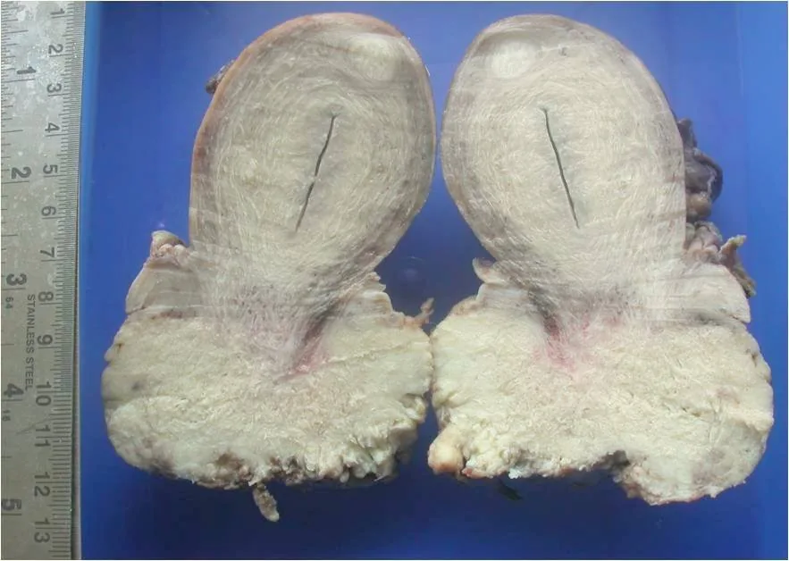 Endoscopy & gross
Definition
- Cervical adenocarcinoma in situ (AIS) is a pre-invasive lesion of the endocervical glandular epithelium and, because of its morphology, is regarded as the precursor of invasive adenocarcinoma.
- Most invasive adenocarcinomas develop from AIS located within or just proximal to the transformation zone.
Classification
Why this special case matters
- Adenocarcinoma arises from the columnar epithelium of the endocervix.
- Cytology therefore detects it less well — it sits endocervically, outside the classic sampling zone.
- The cytology report then carries the suffix g (glandular), e.g. Pap IIIg or IVa-g.
Adenocarcinoma in situ (AIS)
- What sets it apart from CIN: AIS is more often multifocal and discontinuous ("skip lesions").
- Adenocarcinoma is HPV-associated in the majority, especially HPV 18 and 16.
Occurrence & epidemiology
Epidemiology
- About 80–85 % of cervical cancers are squamous cell carcinomas and most of the rest are adenocarcinomas; the proportion of adenocarcinoma has increased over the past two decades.
- Patients with AIS are on average about 5 years younger than those with early invasive adenocarcinoma, reflecting a window of about 5 years between the two.
Aetiopathogenesis
Aetiology and risk factors
- HPV infection is the cause of most cervical cancers; AIS harbours HPV types similar to those found in invasive adenocarcinoma.
- A coexisting squamous neoplasia is common, found in about 55 % of women with AIS in a meta-analysis of 33 studies, reflecting a shared aetiology.
Clinical features
- AIS is almost always asymptomatic and generally not visible on gross examination.
- The lesions lie within the endocervical canal and the glandular clefts and are often multifocal.
Diagnosis
- AIS is usually detected during work-up of an abnormal cervical smear, often actually because of accompanying squamous cell abnormalities.
- Cytology detects glandular lesions poorly, with a sensitivity of only about 45 % in a cited study; adding an HPV test improves detection.
- The colposcopic appearance of AIS is non-specific, and because of their location in the endocervical canal the lesions are difficult to detect colposcopically.
Keep learning in the app
Further reading (selection)
Cross-references
Note: Learning content from the GynFuchs app (flashcards, exam questions, image cases) for medical education – not a treatment recommendation and no substitute for diagnosis or treatment decisions in individual cases. Treatment and management are deliberately not covered on this page.