Hyperprolactinaemia and prolactinoma
Exam relevance: in 8 of 197 board exam reports · rank 82
- Synonyms
- high prolactin, prolactinoma
- Specialty
- Gynaecology · Endocrinology & cycle
- Images
- Histology & cytology 2
- Exam relevance
- 8 of 197 reports · rank 82
- In the app
- 1 flashcards · GynFuchs
- Last updated
- 10/2026 · Dr. Pascal Bafteh
Contents
Images (2)
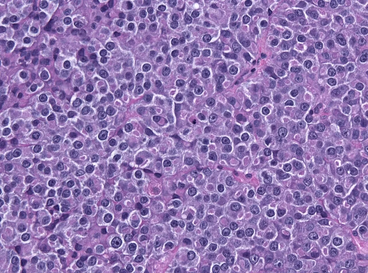 Histology & cytology
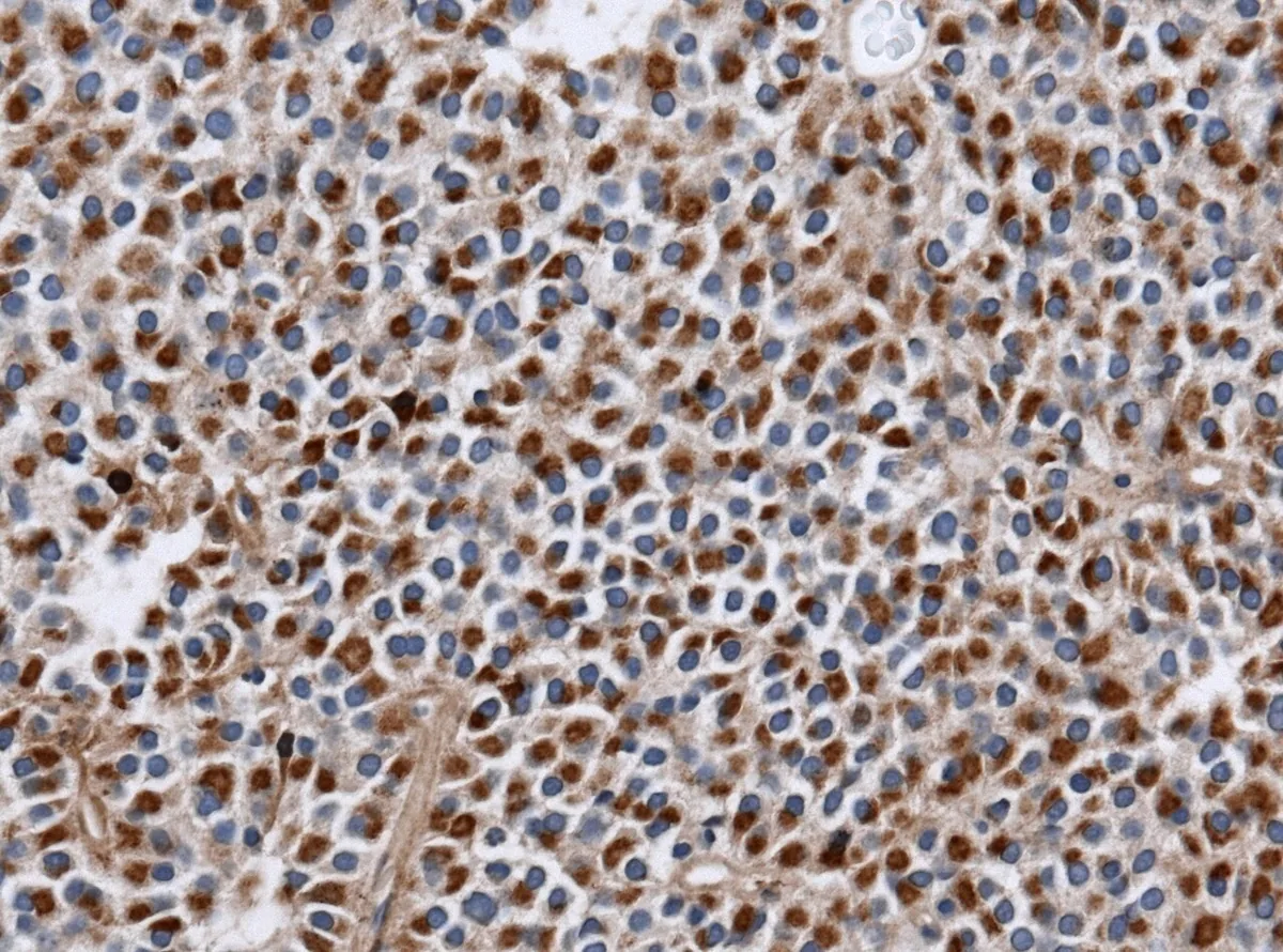 Histology & cytology
Definition
Prolactinoma
- Definition of the size limit: microprolactinoma < 10 mm, macroprolactinoma ≥ 10 mm.
- What is additionally checked for a macroadenoma: visual field (chiasm proximity).
- Hyperprolactinaemia is a raised serum prolactin concentration; a prolactinoma is a benign pituitary adenoma composed of lactotroph cells and the most common hormone-secreting pituitary tumour.
Classification
- By size, microprolactinomas below 10 mm, macroprolactinomas above 10 mm and giant prolactinomas above 4 cm are distinguished.
Occurrence & epidemiology
Epidemiology
- Prolactinomas account for about 40% of all pituitary tumours; between the ages of 20 and 50, women are affected about ten times more often than men, and microadenomas predominate in women.
Aetiopathogenesis
Aetiology and pathogenesis
- Physiological causes include pregnancy, the postpartum period, nipple stimulation, stress, sleep and sexual intercourse.
- Pharmacological causes include dopamine receptor blockers such as risperidone, haloperidol and metoclopramide, antidepressants such as tricyclics and fluoxetine, as well as opioids and oestrogens.
- Besides prolactinoma, pathological causes include lesions of the hypothalamus and pituitary stalk, such as a stalk effect from non-functioning tumours, chronic renal failure, liver cirrhosis and primary hypothyroidism via raised TRH.
Clinical features
Clinical Presentation and Initial Steps
- Typical symptoms: galactorrhoea, cycle disorders/amenorrhoea, infertility.
- Why the cycle is disturbed: suppression of pulsatile GnRH secretion.
- Most common cause in practice: drugs (neuroleptics, MCP, antidepressants).
- Therefore, first: medication history.
- Other causes: hypothyroidism, renal insufficiency, stress.
- Before imaging, one determines: TSH, free thyroid levels, control value.
- In women, galactorrhoea, oligo- or amenorrhoea and infertility predominate; oestrogen deficiency may cause dyspareunia, reduced libido and low bone density up to osteoporosis.
- Space-occupying macroadenomas cause headache, visual field defects, cranial nerve deficits and hypopituitarism; these mass effects predominate mainly in men.
Diagnosis
- Baseline investigations include serum prolactin as well as TSH and T4.
- In prolactinoma, prolactin is typically raised to more than five times normal and correlates with tumour size, whereas non-functioning masses usually do not raise prolactin above three to four times normal.
- Macroprolactin, a prolactin isoform of higher molecular weight and reduced bioactivity, can explain raised values without symptoms; with very large tumours and only modestly raised prolactin, a hook effect can produce falsely low values, revealed by dilution of the sample.
- Contrast-enhanced MRI of the pituitary demonstrates the adenoma, complemented by visual field testing in macroadenomas.
Keep learning in the app
Further reading (selection)
Cross-references
Note: Learning content from the GynFuchs app (flashcards, exam questions, image cases) for medical education – not a treatment recommendation and no substitute for diagnosis or treatment decisions in individual cases. Treatment and management are deliberately not covered on this page.