Endometrioma (chocolate cyst)
Exam relevance: in 12 of 197 board exam reports · rank 59
- Specialty
- Gynaecology · Endometriosis & adenomyosis
- Images
- Ultrasound 1 · Endoscopy & gross 2
- Exam relevance
- 12 of 197 reports · rank 59
- In the app
- 1 flashcards · GynFuchs
- Last updated
- 10/2026 · Dr. Pascal Bafteh
Contents
Images (3)
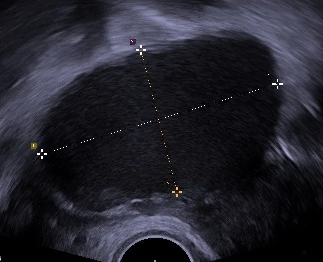 Ultrasound
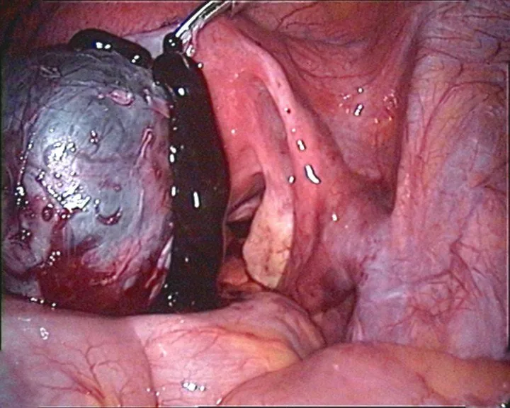 Endoscopy & gross
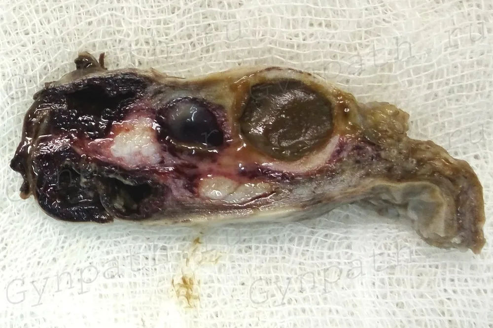 Endoscopy & gross
Definition
- An endometrioma is an endometriotic cyst of the ovary, also called a chocolate cyst because of its old blood content.
- One proposed mechanism is invagination of the ovarian cortex with endometriotic implants that fill the cyst with haemolysed blood.
Classification
- Types include cortical invagination cysts, surface inclusion cyst-related and physiological cyst-related endometriotic cysts, and an unclassified type.
- In endometriosis staging, small endometriomas indicate stage III and large ones stage IV.
Occurrence & epidemiology
Epidemiology and pathogenesis
- Endometriomas occur in 17–44% of patients with endometriosis and account for 35% of all benign ovarian cysts.
- The most accepted theory of endometriosis is implantation of endometrial tissue through retrograde menstruation; the disease is oestrogen-dependent.
Clinical features
- The main manifestations of endometriosis are chronic pelvic pain and impaired fertility; adolescents and, rarely, girls before menarche may also have chronic pelvic pain due to an endometrioma.
- Endometriomas can destroy primordial follicles and thus reduce ovarian reserve; occasionally they rupture, causing acute abdominal pain and peritoneal signs.
- Diagnosis is often delayed, by 8–10 years in some studies.
Histology
- An endometrioma is a pseudocyst with a partial or complete endometrial-like lining.
- The ovarian cortex in the cyst wall contains haemosiderin-laden macrophages and fibrotic components.
Diagnosis
Endometrioma on ultrasound
- On ultrasound, an endometrioma typically appears as homogeneously hypoechoic, smooth-walled cyst.
- On Doppler, there is no internal flow, at most delicate peripheral flow.
- According to IOTA, the endometrioma is benign Easy Descriptor without solid components.
Typical imaging findings
- Ultrasound: Within the ovary lies a rounded cyst whose contents are not anechoic but filled with fine, evenly distributed low-level echoes — the ground-glass appearance. The wall is smooth, with no solid nodules and no papillary components.
- On ultrasound, unilocular or few-loculated cysts (fewer than 5 locules) with homogeneous low-level echoes (ground glass) and poor or absent vascularisation are typical.
- On MRI the shading sign due to old blood products rich in iron and protein is specific; the cysts have higher T1 and lower T2 signal than haemorrhagic cysts.
- Enhancing mural nodules are highly suggestive of malignant transformation, as is loss of the typical shading sign.
Keep learning in the app
Further reading (selection)
Cross-references
More topics: Endometriosis & adenomyosis
Note: Learning content from the GynFuchs app (flashcards, exam questions, image cases) for medical education – not a treatment recommendation and no substitute for diagnosis or treatment decisions in individual cases. Treatment and management are deliberately not covered on this page.