Adenomyosis
Exam relevance: in 17 of 197 board exam reports · rank 39
- Specialty
- Gynaecology · Endometriosis & adenomyosis
- Images
- Ultrasound 2 · Endoscopy & gross 1
- Exam relevance
- 17 of 197 reports · rank 39
- In the app
- 1 flashcards · GynFuchs
- Last updated
- 10/2026 · Dr. Pascal Bafteh
Contents
Images (3)
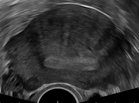 Ultrasound
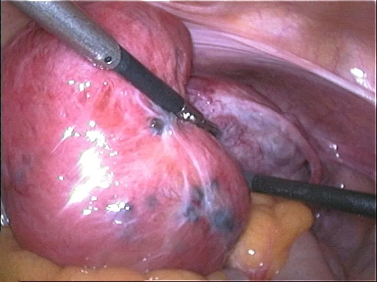 Endoscopy & gross
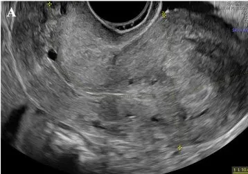 Ultrasound
Definition
What adenomyosis is
- Definition: Adenomyosis is defined as glands/stroma in myometrium with muscle hyperplasia.
- Symptoms: Common symptoms are hypermenorrhoea, dysmenorrhoea, and pressure.
- Association: Adenomyosis often with endometriosis.
- Epidemiology: It predominantly affects perimenopausal women, often after procedures.
Clinical features
More facts from the study questions
- An adenomyoma is characteristically ill-defined, whereas a fibroid has a clear border.
- The MUSA (Morphological Uterus Sonographic Assessment) criteria include myometrial cysts.
- An irregular, thickened or interrupted junctional zone is also a characteristic sonographic finding.
- The pathological definition of adenomyosis is the presence of endometrial-like glands and stroma within the myometrium.
- Adenomyosis can occur in isolation, but it is also frequently associated with endometriosis.
Diagnosis
MUSA
- Acronym: MUSA stands for Morphological Uterus Sonographic Assessment.
- Uterine shape: A MUSA criterion is globular uterus with asymmetric walls.
- Cysts: Another sonographic criterion is myometrial cysts.
- Echogenicity: The MUSA criteria also include hyperechoic islands and fan-shaped shadowing.
- Junctional zone: A typical finding is an irregular, thickened junctional zone.
- Vascularisation: The vessel architecture shows translesional, penetrating vessels.
Typical imaging findings
- Ultrasound: The uterus is globularly enlarged with asymmetric anterior and posterior wall thickness. Fine, fan-shaped shadow stripes run vertically through the myometrium — the typical venetian-blind pattern; the endometrium itself is normal.
- Ultrasound: The myometrium is asymmetrically thickened and studded with small anechoic spaces. The junction between endometrium and myometrium is ill-defined and wavy, without any encapsulated mass.
Keep learning in the app
Further reading (selection)
Cross-references
More topics: Endometriosis & adenomyosis
Note: Learning content from the GynFuchs app (flashcards, exam questions, image cases) for medical education – not a treatment recommendation and no substitute for diagnosis or treatment decisions in individual cases. Treatment and management are deliberately not covered on this page.