Endometriosis
Exam relevance: in 37 of 197 board exam reports · rank 17
- Specialty
- Gynaecology · Endometriosis & adenomyosis
- Images
- Endoscopy & gross 11 · Ultrasound 1
- Exam relevance
- 37 of 197 reports · rank 17
- In the app
- 6 flashcards · GynFuchs
- Last updated
- 10/2026 · Dr. Pascal Bafteh
Contents
Images (12)
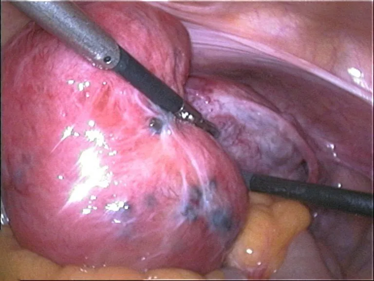 Endoscopy & gross
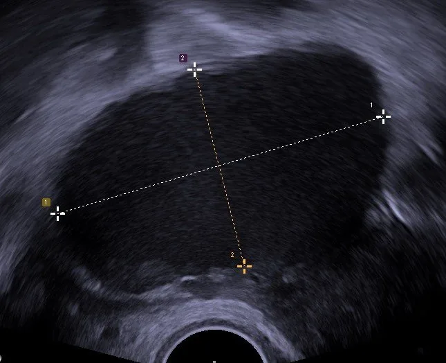 Ultrasound
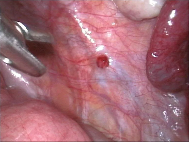 Endoscopy & gross
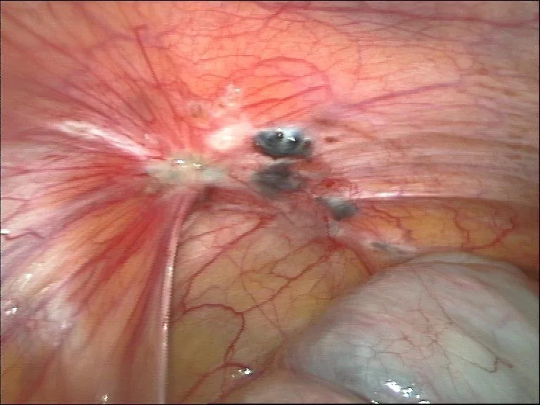 Endoscopy & gross
 Endoscopy & gross
 Endoscopy & gross
 Endoscopy & gross
 Endoscopy & gross
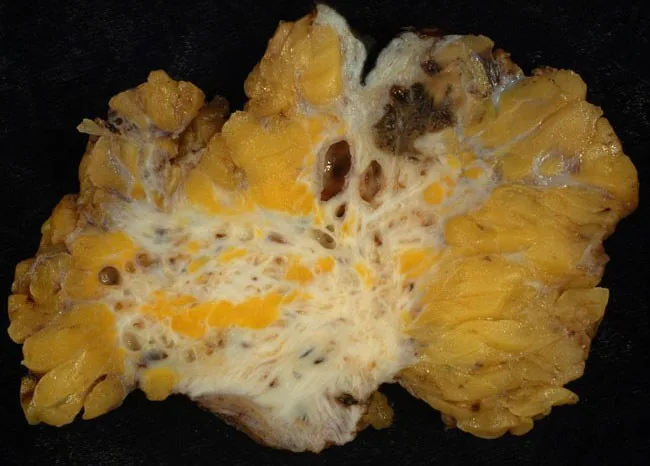 Endoscopy & gross
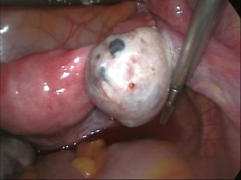 Endoscopy & gross
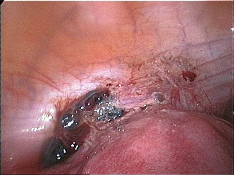 Endoscopy & gross
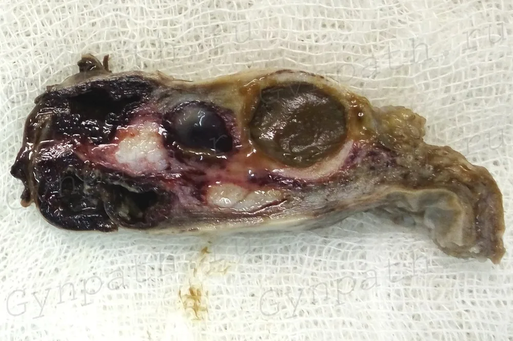 Endoscopy & gross
Definition
- Endometriosis is defined as ectopic endometrium-like tissue.
- Adenomyosis is defined as endometrial tissue in the myometrium.
- Affected are 10% of women (fertile), 50% with chronic pain.
- The condition is a chronic, oestrogen-dependent disease.
Classification
RASRM
- The abbreviation rASRM stands for rev. classification of the Am. Society for Repr. Medicine.
- According to rASRM, what is assessed is the peritoneal/ovarian lesions, adhesions, Douglas pouch.
- The point score results in four stages (minimal to severe).
- The weakness of the rASRM classification is that it doesn't map DIE/organ involvement/pain.
#ENZIAN
- The #ENZIAN classification describes compartment-based, including deep infiltrating form.
- Compartment P stands for peritoneal endometriosis.
- Compartments O and T stand for ovaries; T: tubo-ovarian junction.
- Compartments A, B, and C stand for vagina/rectovaginal space, sacrouterine lig., rectum.
- The suffix F (for “far”) denotes A: adenomyosis, B: bladder, U: ureter, I: bowel, O: other.
- The size of the lesions is coded in 3 grades: <1 cm, 1–3 cm, >3 cm.
Aetiopathogenesis
Theories
- Sampson's transplantation theory states endometrial cells reach the abdomen retrogradely.
- Meyer's metaplasia theory states coelomic epithelium transforms into endometrial tissue.
- The archimetra theory explains adenomyosis via disturbed uterine peristalsis.
- For extragenital spread, lymphogenous and hematogenous spread is assumed.
- As no single theory explains all phenomena, it is assumed to be a multifactorial process with immune and genetic factors.
Risk and Protective Factors
- The risk of endometriosis is increased by early menarche, short cycles, nulliparity, family history.
- Outflow obstructions such as a vaginal septum increase the risk because they increase retrograde menstruation.
Why Fertility is Impaired
- Mechanical disturbances: Fertility is mechanically impaired by adhesions, tube/ovary displacement, Douglas obliteration.
- Functional disturbances: Functional disturbances include peritoneum, oocyte quality, implantation.
- Adenomyosis: In adenomyosis, there is the additional factor of disturbed junctional zone, altered uterine peristalsis.
- Time to pregnancy: About one in two to three women with endometriosis reports an increased time to pregnancy.
Clinical features
Four Ds
- The first D of the cardinal symptoms is dysmenorrhoea, i.e., pain with menstrual bleeding.
- The second D is dyspareunia.
- The third D is dyschezia, i.e., pain during defecation.
- The fourth D is dysuria.
- This series of cardinal symptoms is often supplemented by infertility as a fifth cardinal symptom.
Diagnostic Latency
- Between the first symptoms and the final diagnosis, there are on average years.
- A main reason for the diagnostic latency is the trivialisation of menstrual pain.
- There is no correlation between extent of lesions and severity of pain.
Locations
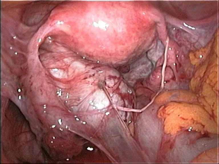
- Most commonly affected are the peritoneum, ovaries, sacrouterine ligg., pouch of Douglas.
- Genital endometriosis can also affect the rectovaginal septum, vagina, cervix.
- Extragenitally, lesions occur in the bowel, bladder, ureter, umbilicus, diaphragm, pleura.
- Abdominal wall endometriosis typically develops in a C-section scar with cyclical pain.
Appearance of Lesions
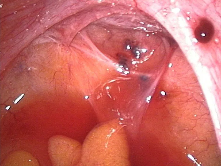
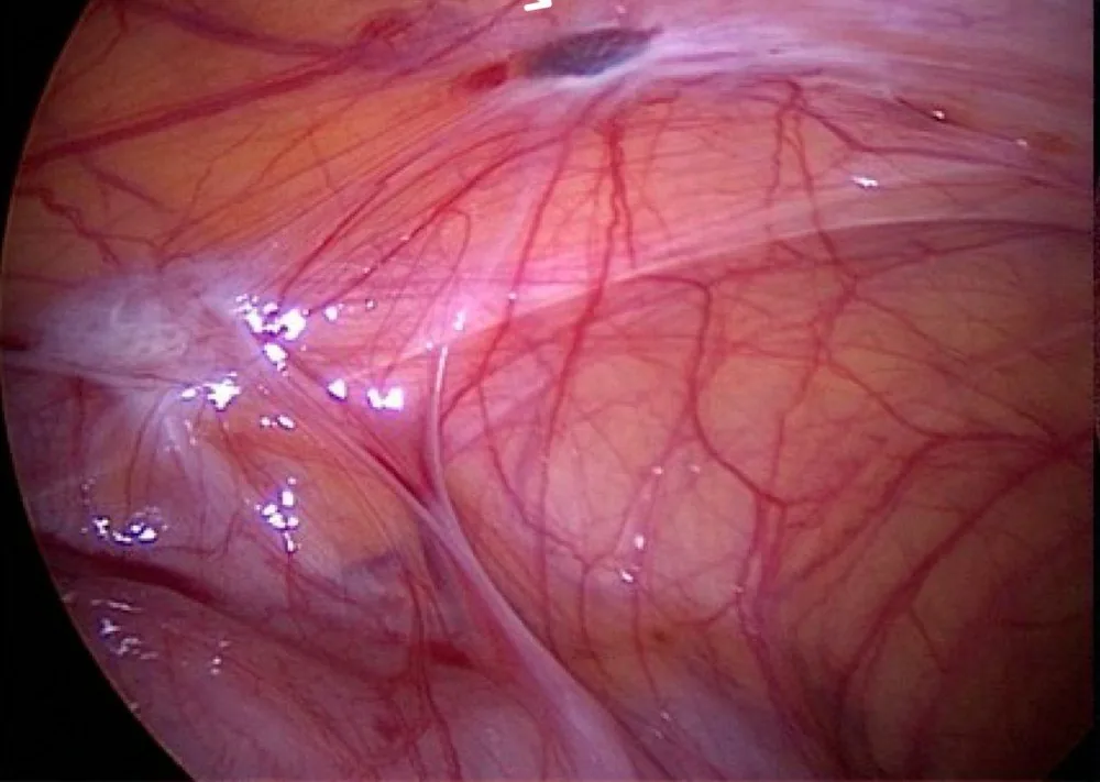
- Fresh, active lesions are red and well-vascularised.
- Older lesions are black-blue as powder-burn lesions.
- Burnt-out lesions are white, fibrotic with stellate scar retraction.
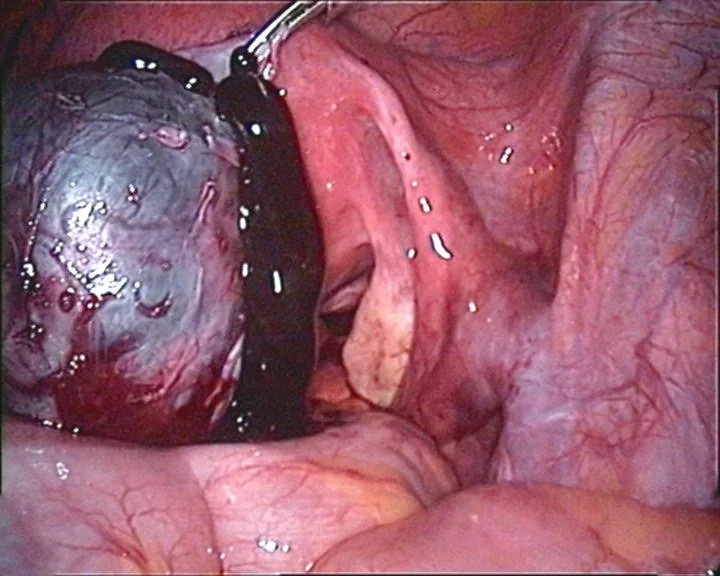
- An endometriotic cyst of the ovary is called an endometrioma (chocolate cyst).
More facts from the study questions
- The four 'D's of the cardinal symptoms are dysmenorrhoea, dyspareunia, dyschezia and dysuria.
- Dyspareunia is pain during sexual intercourse.
- There is no correlation between the extent of the lesions (stage) and the severity of pain.
- The patient's level of suffering is a central aspect of the medical history.
- It is assessed by analgesic use and limitations in daily life, among other factors.
- Endometriosis is the presence of endometrium-like tissue outside the uterine cavity.
- Adenomyosis is the specific form with this tissue present within the myometrium.
- Endometriosis is a chronic, oestrogen-dependent disease.
- With the fall in oestrogen levels in the postmenopause, the symptoms cease.
- Sampson's transplantation theory is one of the central hypotheses on the origin of endometriosis.
Diagnosis
Inspection and Speculum Examination
- Speculum examination: Using a bivalve speculum, one looks for bluish-red lesions in the posterior vaginal fornix.
- Diagnostics for suspected TIE: If there is suspicion of deeply infiltrating endometriosis, a targeted biopsy under vision is possible.
- Examination of the abdominal wall: During inspection, c-section scar for cyclical pain.
Palpation
- Bimanual examination: One palpates bimanually for size, position, mobility, and tenderness of the uterus.
- Finding in TIE: A bimanually fixed, retroverted uterus is suggestive of for DIE with adhesions.
- Rectovaginal examination: This is performed to to palpate for firm, painful nodules.
- Optimal timing for examination: The examination is most informative during/shortly after menstruation.
Role of Diagnostics
- Gold standard: The gold standard for confirming the diagnosis remains laparoscopy with histology.
- Laboratory diagnostics: In terms of laboratory tests, there is no specific laboratory marker.
Systematic Assessment
- The sliding sign tests mobility of uterus against rectum.
- A negative sliding sign means an obliteration of the pouch of Douglas by adhesions.
- A systematic search is also conducted for nodules on septum, ligaments, bladder & bowel wall.
MRI and Limitations
- MRI is superior to sonography for deep endometriosis with bowel/bladder involvement.
- Imaging generally fails to detect purely peritoneal endometriosis.
- In case of bowel involvement, colonoscopy/endorectal ultrasound for stenosis may also be considered.
Keep learning in the app
Further reading (selection)
Cross-references
More topics: Endometriosis & adenomyosis
Note: Learning content from the GynFuchs app (flashcards, exam questions, image cases) for medical education – not a treatment recommendation and no substitute for diagnosis or treatment decisions in individual cases. Treatment and management are deliberately not covered on this page.