Deep infiltrating endometriosis
Exam relevance: in 6 of 197 board exam reports · rank 93
- Specialty
- Gynaecology · Endometriosis & adenomyosis
- Images
- Endoscopy & gross 2
- Exam relevance
- 6 of 197 reports · rank 93
- In the app
- 1 flashcards · GynFuchs
- Last updated
- 10/2026 · Dr. Pascal Bafteh
Contents
Images (2)
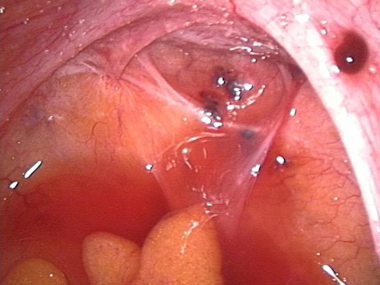 Endoscopy & gross
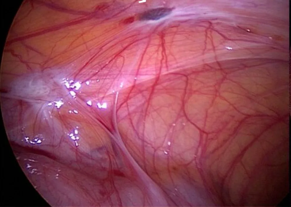 Endoscopy & gross
Definition
- Deep infiltrating endometriosis is present when endometriotic tissue with fibrosis and hyperplasia infiltrates more than 5 mm beneath the peritoneum; lesions under 5 mm are regarded as superficial.
- Endometriosis is characterised by endometrial glands and stroma outside the uterine cavity and is an oestrogen-dependent inflammatory disease.
Classification
#ENZIAN classification
- The #ENZIAN classification describes compartment-based, including deep infiltrating form.
- Compartment P stands for peritoneal endometriosis.
- Compartments O and T stand for ovaries; T: tubo-ovarian junction.
- Compartments A, B, and C stand for vagina/rectovaginal space, sacrouterine lig., rectum.
- The suffix F (for “far”) denotes A: adenomyosis, B: bladder, U: ureter, I: bowel, O: other.
- The size of the lesions is coded in 3 grades: <1 cm, 1–3 cm, >3 cm.
Sites
- In endometriosis overall, the ovaries are most commonly involved, followed by the posterior broad ligament, the anterior and posterior cul-de-sac and the uterosacral ligaments; endometriotic nodules can also affect the bowel, ureter, bladder and urethra.
Occurrence & epidemiology
Epidemiology
- The prevalence of endometriosis is reported as about 10–18% in women of reproductive age and 20–50% in infertile women; the deep infiltrating form accounts for 15–30% of diagnosed cases.
Clinical features
- Typical are dysmenorrhoea, chronic pelvic pain and infertility.
- Depending on the site, deep dyspareunia, dyschezia or rectal bleeding during menses, and with bladder involvement dysuria and haematuria, may occur.
- On examination a thickened or nodular rectovaginal septum or nodules on the uterosacral ligaments may be palpable, although findings may also be normal.
Diagnosis
- In a meta-analysis, physical examination reached a pooled sensitivity of 71% and specificity of 69%, transvaginal ultrasound 76% and 94%, and MRI 82% and 87%.
- Additional techniques such as assessing pelvic organ mobility (sliding sign) and rectal water contrast improve the diagnostic performance of transvaginal ultrasound.
Keep learning in the app
Further reading (selection)
- Comparison of physical examination, ultrasound techniques and magnetic resonance imaging for the diagnosis of deep infiltrating endometriosis: A systematic review and meta-analysis of diagnostic accuracy studies (Exp Ther Med 2020, PubMed Central)
- StatPearls: Endometriosis
- MSD Manual Professional: Endometriosis
- DocCheck Flexikon, Endometriose
Cross-references
More topics: Endometriosis & adenomyosis
Note: Learning content from the GynFuchs app (flashcards, exam questions, image cases) for medical education – not a treatment recommendation and no substitute for diagnosis or treatment decisions in individual cases. Treatment and management are deliberately not covered on this page.