Shock (types of shock)
Board exam relevance: in 4 of 105 exam reports · rank 90- Synonyms
- circulatory shock, circulatory failure, septic shock, cardiogenic shock, hypovolaemic shock
- Specialty
- Internal medicine · Critical care & emergency medicine
- Images
- CT 1
- Last updated
- 10/2026 · Dr. Pascal Bafteh
Contents
Images (1)
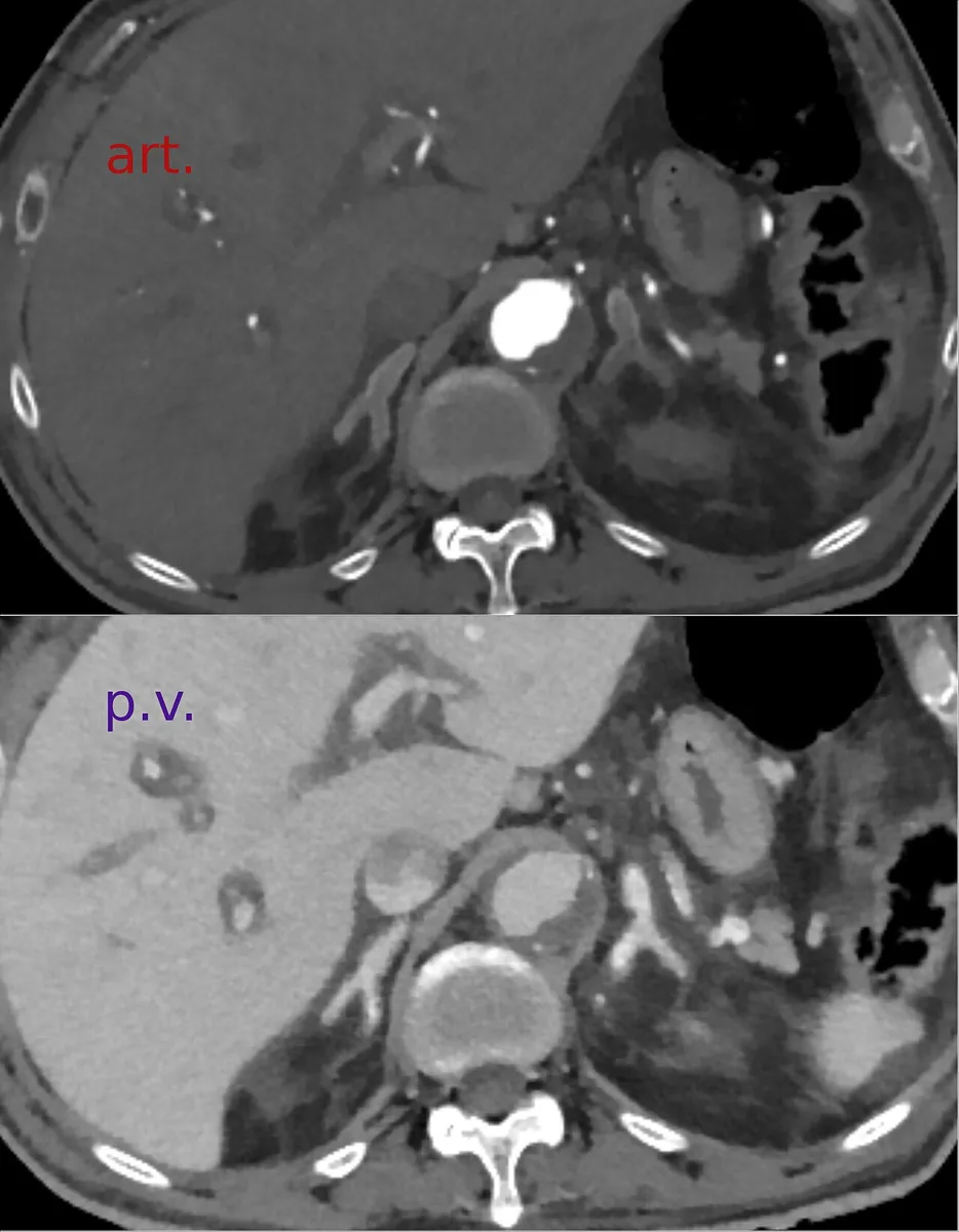 CT
CTDefinition
Shock is tissue hypoperfusion due to acute circulatory failure with inadequate oxygen delivery and resulting cellular dysfunction and damage. It is defined by symptoms and signs of hypoperfusion: altered mental status, cool, clammy or cyanotic skin, reduced urine output and a raised lactate (above 2 mmol/l).
Hypotension is common but not synonymous with shock: not every person in shock is hypotensive, and not every hypotension means shock. What matters is impaired oxygen utilisation at the cellular level.
Classification
By mechanism, four types of shock are distinguished, which occur alone or in combination:
- Hypovolaemic shock: critical decrease in intravascular volume; reduced venous return (preload) lowers ventricular filling and stroke volume
- Distributive shock: relative volume deficit due to dilatation of arteries or veins with normal blood volume; this includes septic, anaphylactic and neurogenic shock as well as shock caused by certain drugs or poisons
- Cardiogenic shock: relative or absolute reduction in cardiac output due to a primary cardiac disorder
- Obstructive shock: mechanical interference with filling or emptying of the heart or great vessels
According to Sepsis-3, septic shock is sepsis with persistent hypotension in which a mean arterial pressure of at least 65 mmHg can only be maintained with vasoactive drugs, and a lactate above 2 mmol/l despite adequate fluid volume.
Aetiopathogenesis
- Hypovolaemic: bleeding (hemorrhagic shock), e.g. from trauma, peptic ulcer, esophageal varices, ruptured aortic aneurysm or ruptured ectopic pregnancy; fluid losses via the skin (burns, heavy sweating), the gut (vomiting, diarrhea) or the kidneys (e.g. diabetes mellitus, AVP deficiency, polyuric phase after kidney injury, adrenal insufficiency); fluid shift into the tissues in inflammation, sepsis, bowel ischemia or acute pancreatitis; inadequate fluid intake
- Distributive: sepsis, anaphylaxis, toxic shock syndrome, severe spinal cord injury usually above T4 (neurogenic shock), certain drugs or poisons
- Cardiogenic: myocardial ischemia and infarction, myocarditis, arrhythmias (tachycardia or bradycardia), acute mitral or aortic regurgitation, ventricular septal rupture, prosthetic valve malfunction
- Obstructive: tension pneumothorax, cardiac tamponade, compression of the venae cavae, atrial tumor or thrombus, pulmonary embolism
Pathophysiology: when oxygen delivery falls, tissues initially extract more oxygen. Low blood pressure activates the sympathetic nervous system with vasoconstriction and tachycardia; blood flow is redistributed in favour of the heart and brain (centralisation). Cortisol, aldosterone, renin and glucose are also released. Once oxygen is insufficient for aerobic metabolism, lactate rises. In hypoperfused tissues the inflammatory and clotting cascades are activated, up to disseminated intravascular coagulation. Direct injury and reperfusion injury can trigger multiple organ dysfunction syndrome (MODS) – progressive failure of at least two organs, most commonly in sepsis.
Clinical features
- Altered mental status: lethargy, confusion, somnolence
- Skin: hands and feet pale, cool, clammy and often cyanotic, as are earlobes, nose and nail beds
- Pulse: weak and usually rapid; often only femoral or carotid pulses are palpable
- Capillary refill time: prolonged (over 2 seconds) in hypovolaemic, cardiogenic and obstructive shock; often normal in distributive shock, with warm, flushed skin and possibly a bounding pulse
- Compensatory signs: sweating, tachycardia, tachypnea
- Vital signs: there are no absolute cut-offs; common are heart rate above 100/min, respiratory rate above 22/min and systolic blood pressure below 90 mmHg or a fall of 30 mmHg from baseline
- Oliguria
Clues to the cause: fever and chills in septic shock, urticaria or wheezing in anaphylactic shock, distended neck veins, muffled heart sounds and pulsus paradoxus in cardiac tamponade.
Organ consequences: acute respiratory distress syndrome (ARDS), acute tubular necrosis with kidney failure, reduced cardiac output and arrhythmias, ileus and mucosal bleeding in the gut, hepatocellular necrosis with rising transaminases and bilirubin, coagulation disorders.
Diagnosis
The diagnosis is based mainly on the history (e.g. febrile illness, injury with bleeding) and signs of hypoperfusion and compensation. The initial assessment follows the ABCDE approach; the level of consciousness is assessed with the Glasgow Coma Scale.
Laboratory tests:
- lactate: above 2 mmol/l indicates tissue hypoperfusion
- urea, creatinine and electrolytes (kidney injury, metabolic disturbances)
- liver tests (liver injury)
- full blood count (blood loss, infection)
- coagulation including fibrinogen (disseminated intravascular coagulation)
- blood gas analysis with pH, base deficit, pCO₂ and pO₂
- depending on the suspected cause, cardiac biomarkers, blood and urine cultures, pregnancy test, amylase and lipase
Instrumental diagnostics:
- echocardiography to assess cardiac output
- chest X-ray
- bedside (point-of-care) ultrasound to assess cardiac filling, cardiac function and volume status
- ECG; further imaging (e.g. CT) depending on the suspected cause
No single finding is diagnostic on its own; trends and the overall clinical picture are decisive. Identifying the cause is often more important than classifying the type of shock.
Keep learning in the app
Further reading (open access)
Cross-references
More topics: Critical care & emergency medicine
Note: Learning content for medical education – not a treatment recommendation and no substitute for diagnosis or treatment decisions in individual cases. Treatment and management are deliberately not covered on this page.